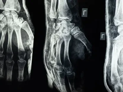

Modern diagnostic imaging often faces a significant hurdle when the patient is much smaller than the machines designed to examine them, particularly within the sensitive environment of a magnetic resonance imaging suite. While adult patients benefit from standardized equipment, infants and young children frequently endure suboptimal scans because existing radio-frequency coils are rigid and oversized. This physical mismatch leads to a significant loss in signal sensitivity, as the gap between the child’s body and the sensor introduces electronic noise that degrades the final image quality. Furthermore, the necessity of keeping a child perfectly still within a cavernous machine adds another layer of complexity to an already stressful situation for families. Standard hardware simply lacks the ergonomic flexibility to contour to the unique anatomical curves of a fragile neonate. This technological gap creates a disparity in medical care, as the precision of data-driven medicine becomes hindered by the physical constraints of legacy hardware. The quest for a solution has led researchers to look beyond traditional manufacturing, seeking ways to create sensors that are as dynamic and adaptable as the patients they are intended to serve, ensuring that every scan provides the clarity needed for life-saving interventions.
Engineering Adaptive Solutions: Conforming Technology to the Patient
Material Innovation: The Science of Stretchable Conductive Polymers
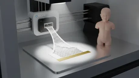
The introduction of 3D-printed MRI sensors marks a pivotal shift toward patient-centric hardware, utilizing advanced additive manufacturing to bridge the gap between human anatomy and electronic diagnostic tools. Researchers have pioneered a method to print conductive elements onto stretchable, polymer-based substrates that behave more like a second skin than a piece of medical equipment. These flexible coils can be wrapped directly around a limb, the torso, or even the delicate head of a newborn, ensuring that the sensor maintains constant contact with the skin surface. This proximity is vital because the strength of the magnetic signal diminishes exponentially as the distance from the target increases. By eliminating the air gaps typical of rigid, adult-sized sensors, this new manufacturing technique allows for an unprecedented capture of biological data. The result is a highly sensitive diagnostic interface that conforms to the patient, rather than forcing the patient to conform to the machine. This material breakthrough ensures that children no longer have to be subjected to the discomfort of bulky, ill-fitting arrays that were never designed for their small frames.
In traditional pediatric imaging, the oversized nature of coils often results in images that are too grainy for certain complex diagnoses, such as identifying minute structural defects in a developing heart. By utilizing skin-tight 3D-printed electronics, radiologists can now achieve high-contrast visuals that were previously unattainable with standard equipment. This clarity is essential for detecting early-stage neurological issues or subtle abdominal pathologies that might otherwise go unnoticed until they become more severe. The ability to generate such precise data ensures that medical teams can make informed decisions with greater confidence. This transition to high-resolution customized sensors represents a fundamental improvement in the diagnostic capabilities of modern hospitals, prioritizing accuracy for the most vulnerable. Moreover, the enhanced resolution significantly reduces the need for repeated scans, which minimizes the time a young child must spend under sedation or within the high-stress environment of the MRI room. By 2027, this approach is expected to become the gold standard for all specialized neonatal intensive care units.
Rapid Customization: Scaling Hardware to the Growth Lifecycle
One of the most compelling aspects of this additive manufacturing breakthrough is the efficiency and cost-effectiveness it brings to the specialized field of pediatric medical hardware. Historically, creating custom-fitted components for medical imaging was a prohibitively expensive and time-consuming endeavor, often requiring weeks of specialized labor and thousands of dollars in investment. The current 3D-printing process has revolutionized this timeline, allowing a personalized coil to be produced in roughly ten minutes. This rapid turnaround time means that a medical facility can print a sensor tailored to a child’s specific body measurements while the family is still in the clinic, streamlining the entire diagnostic workflow. Furthermore, the material costs have plummeted, with each customized sensor now costing approximately thirty dollars to manufacture. This reduction in overhead makes it feasible for hospitals to maintain a dynamic inventory of custom tools that can be discarded or recycled responsibly. This agility allows the medical field to keep pace with the urgent needs of emergency diagnostics without the burden of excessive costs.
The dynamic nature of childhood growth presented a unique challenge for medical practitioners who had to monitor chronic conditions over several months or years. A sensor that fitted a newborn perfectly would quickly become undersized as the infant developed, necessitating a new solution at every stage of the growth cycle. The digital workflow associated with 3D printing allowed medical technicians to simply scale a digital model and print a larger version as the patient grew. This adaptability ensured that the quality of imaging remained consistent throughout the entire course of a child’s treatment, providing a seamless stream of data that reflected the patient’s physical evolution. Instead of relying on a limited selection of standard sizes, doctors ordered precise adjustments that accounted for growth spurts or specific anatomical changes. This level of customization provided a continuous and accurate medical history, allowing for long-term health monitoring that was tailored to specific anatomical needs. Hospitals that adopted these protocols from 2026 to 2028 saw a marked increase in the speed of diagnostic transitions, successfully bridging the gap between neonatal care and early childhood medicine.