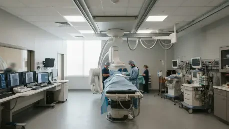

The conventional landscape of oncology is undergoing a profound structural shift as nuclear medicine transitions from a secondary diagnostic tool to the very cornerstone of personalized therapeutic strategies. This evolution is driven by the seamless integration of high-resolution molecular imaging with targeted radioligand therapies, allowing clinicians to visualize the unique biochemical signature of a patient’s malignancy before a single drop of medicine is administered. By 2026, the adoption of these precision protocols has moved beyond experimental settings, becoming a standard of care that replaces the “one-size-fits-all” approach that once dominated cancer treatment. These advancements provide a window into the cellular level of disease, enabling doctors to predict how a tumor will react to specific isotopes and adjust dosages with unprecedented accuracy. This level of granularity not only improves the odds of a successful remission but also ensures that patients are not subjected to the toxic side effects of treatments that their specific tumor biology would render ineffective.
Enhancing Precision in Prostate Cancer Management
The primary frontier of this revolution is the management of prostate cancer, where the synergy between PSMA PET/CT and multiparametric MRI has redefined clinical pathways. In 2026, the identification of candidates for focal therapy—a treatment designed to destroy the tumor while sparing healthy prostatic tissue—requires a level of precision that traditional biopsy cannot provide. Recent research confirms that using these dual imaging modalities in tandem significantly enhances the detection of multifocal disease and extracapsular extension, which are critical factors that dictate the success of localized interventions. This multilayered screening process acts as a safeguard, ensuring that only those with truly localized disease undergo focal treatment. Furthermore, large-scale clinical analyses involving thousands of patients demonstrate that utilizing PSMA PET/CT for initial staging correlates directly with superior survival outcomes, particularly for high-risk individuals who require highly targeted radiation therapy to prevent recurrence and improve long-term health.
Monitoring the efficacy of radioligand therapy presents its own set of unique challenges, especially when traditional biomarkers like PSA levels do not align with visual evidence of disease. Research has highlighted a phenomenon known as “discordance,” where nearly half of the patient population might show stable blood tests despite imaging revealing localized tumor progression in the bones or lymph nodes. This discrepancy emphasizes the necessity of shifting away from a reliance on simple blood work in favor of regular serial imaging to truly understand treatment response. When standard PSMA scans fail to pinpoint the source of a recurrence despite rising PSA levels, secondary tracers such as 18F-fluciclovine have emerged as an essential diagnostic fallback. This specific tracer has successfully identified hidden recurrences in over half of patients with negative PSMA scans, leading to immediate changes in clinical management for the vast majority. By 2026, this layered diagnostic strategy ensures that no trace of the disease remains unaddressed or untreated.
Expanding Theranostics and Improving Patient Quality of Life
The concept of “theranostics” is reaching its full potential through the use of Fibroblast Activation Protein (FAP) imaging, which bridges the gap between diagnostic insight and therapeutic action. Diagnostic FAPI PET scans are now being used to predict the exact radiation dosimetry a tumor will receive during subsequent therapy, allowing physicians to calculate the specific absorption rate for solid tumors with high reliability. This predictive capability is particularly transformative for lung cancer patients, where PET imaging of PD-L1 expression can determine the likelihood of a positive response to immunotherapy. Unlike a physical biopsy, which only captures a tiny fraction of a tumor’s biological makeup, molecular imaging provides a noninvasive map of the entire body’s disease burden. This whole-body perspective is vital in 2026, as it identifies which patients will truly benefit from complex immune-based treatments. By screening out those who would only experience toxicity without the benefit, nuclear medicine is optimizing healthcare resources and sparing patients from unnecessary physiological strain.
Clinicians and researchers have moved beyond a narrow focus on tumor eradication to prioritize the functional health and long-term quality of life for those undergoing intensive cancer care. This holistic approach is demonstrated in recent findings regarding gallbladder function, where nuclear imaging identified “hypercontractility” as a significant cause of patient distress, leading to effective surgical solutions for a previously underserved group. Simultaneously, the field addressed the debilitating side effects of radiopharmaceutical treatments, such as the salivary gland damage that often follows PSMA therapy. Innovative techniques involving localized injections of botulinum toxin and protective patches successfully reduced the uptake of radioactive ligands in healthy tissue by significant margins. These interventions ensured that the pursuit of a cure did not permanently diminish a patient’s daily comfort or oral health. Moving forward, the industry has established a framework where diagnostic accuracy and symptom mitigation are treated as inseparable, paving the way for a healthcare model where precision is defined by both the elimination of disease and the preservation of human well-being.