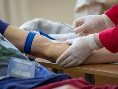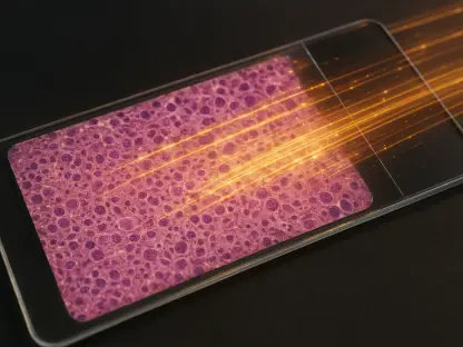

Traditional liver biopsies often fail to capture the full picture of organ health, leading to sampling errors that can result in the unnecessary discard of viable donor livers. This invasive procedure provides only a microscopic glimpse into a large and complex organ, often missing localized pathologies that affect overall function. In the current medical landscape of 2026, technology has pivoted toward non-invasive alternatives that utilize artificial intelligence to analyze high-resolution imaging with unprecedented precision. These AI-driven tools do not just look at the surface; they delve into the cellular density and fat distribution across the entire liver, offering a comprehensive assessment that a needle biopsy cannot replicate. As the global demand for transplantable organs outpaces supply, the need for more accurate diagnostic methods has never been more urgent. By integrating sophisticated software into existing radiology workflows, clinicians now see a clearer pathway toward determining tissue viability.
The Evolution: Algorithmic Liver Assessment
The core of this transformation lies in the application of Convolutional Neural Networks trained on vast datasets of histological and radiological images. These systems excel at identifying subtle patterns in hepatic steatosis and fibrosis that are often invisible to the naked eye of even the most experienced pathologists. Unlike manual grading, which relies on subjective visual estimation, AI-enhanced imaging provides a quantitative metric that standardizes the assessment process across different medical facilities. This consistency ensures that a liver evaluated in one region meets the same rigorous standards as one in another, effectively eliminating the variability that plagues human-led diagnostics. Furthermore, modern platforms are now capable of performing real-time analysis during the imaging session itself, allowing surgeons to make critical decisions within minutes. This speed is vital in transplant scenarios where every hour counts toward the success of the life-saving procedure.
Beyond simple pattern recognition, these advanced algorithms facilitate a three-dimensional reconstruction of the liver’s vascular and parenchymal structure. While a needle biopsy samples less than one-fifty-thousandth of the total organ, AI-enhanced computed tomography and magnetic resonance imaging assess the entire volume. This shift from a localized sample to a holistic analysis addresses the issue of spatial heterogeneity, where disease may be present in one lobe but absent in another. By mapping the distribution of fat and scarring throughout the whole organ, clinicians can identify specific zones of concern that a traditional biopsy would likely bypass. This depth of insight allows for a more nuanced understanding of liver health, particularly in cases of non-alcoholic fatty liver disease. The ability to monitor the progression or regression of such conditions through repeat imaging, without the trauma of surgery, represents a significant milestone in modern clinical hepatology.
Clinical Integration: Optimizing Safety and Supply
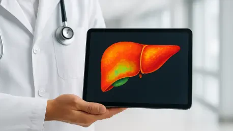
The practical application of this technology has already shown remarkable results in the field of organ transplantation, where efficiency directly translates to lives saved. High-resolution scans processed by AI can identify livers previously categorized as high-risk or marginal due to suspected steatosis, revealing that many are actually within safe parameters for transplantation. This capability has effectively increased the pool of available organs by reducing the discard rate of healthy tissue that appeared problematic on conventional imaging. In several recent clinical trials, the implementation of AI-assisted screening reduced the time required for donor evaluation significantly. Moreover, the accuracy of these automated assessments has matched or exceeded the performance of traditional biopsies in predicting post-transplant function. By providing a reliable method for evaluation, medical centers are optimizing their resource allocation and ensuring that every viable organ reaches a recipient in need.
To fully realize the potential of AI-enhanced imaging, stakeholders prioritized the standardization of data sharing protocols across international medical networks. It became clear that the success of these diagnostic tools depended on the diversity and volume of the data used for training. Health organizations moved to establish unified benchmarks for algorithm performance, ensuring that every software update maintained clinical safety and diagnostic accuracy. Practitioners were encouraged to undergo specialized training to interpret AI-generated insights alongside traditional radiological data, fostering a collaborative relationship between human expertise and machine precision. Future efforts focused on expanding these technologies to mobile imaging units, which allowed for rapid assessment in remote areas. By shifting the focus toward a data-centric diagnostic model, the medical community successfully mitigated the limitations of physical sampling, ensuring the transition was supported by a robust framework of validation.