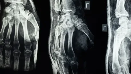

The diagnostic precision of modern oncology often rests on the ability to detect skeletal metastases before they become visible on standard x-rays, yet the visual complexity of bone scintigraphy has long remained a barrier to automated interpretation. Standard artificial intelligence models trained on everyday images like cats or cars fail to accurately identify the subtle grayscale signals and high noise levels inherent in functional nuclear medicine. While technologies like Computed Tomography (CT) and Magnetic Resonance Imaging (MRI) provide crisp anatomical structures, Single-Photon Emission Computed Tomography (SPECT) focuses on physiological activity, resulting in low-resolution images where anomalies appear as faint, blurred regions of tracer uptake. A recent breakthrough presented in the journal Applied Intelligence marks a shift in how these functional scans are processed, introducing a deep learning framework designed to bridge the gap between grainy pixels and the high-fidelity clinical language used by radiologists.
Advanced Architectural Design for Medical Imaging
Visual Feature Extraction and Signal Recalibration
A foundational hurdle in medical image analysis is that most computer vision algorithms lack the specific context required to interpret nuclear medicine data. The researchers addressed this by developing the Domain-Adaptive Visual Feature Extractor (DAVFE), a component that bypasses the limitations of generic training datasets. By utilizing a self-supervised contrastive pretraining strategy on a vast library of unlabeled SPECT scans, the model learns to recognize the fundamental textures and patterns unique to radioactive tracer distribution. This process allows the system to internalize the variance in bone density and metabolic activity without requiring an impractical amount of manually labeled data from the start. Consequently, the AI develops a baseline sensitivity to the low-contrast environment of scintigraphy, enabling it to distinguish between the inherent noise of the imaging process and the critical physiological signals that indicate potential skeletal pathology in a clinical setting.
Beyond just identifying patterns, the framework employs an attention-based recalibration mechanism to fine-tune its visual perception. This technical layer functions as a sophisticated filter that isolates the areas of highest clinical significance within a scan, specifically focusing on hotspots where tracer uptake is abnormally concentrated. In traditional nuclear medicine, background noise often obscures these vital signs, leading to potential misinterpretation or overlooked lesions. The attention mechanism effectively suppresses irrelevant visual data while amplifying the subtle fluctuations in grayscale intensity that a human radiologist would prioritize. By mimicking this human-like focus, the system ensures that the reporting engine remains anchored to the most relevant anatomical findings. This recalibration is not merely about image enhancement; it is about directing the model’s computational resources toward the regions that ultimately dictate a patient’s diagnosis and treatment plan for 2026 and beyond.
Semantic Alignment and Anatomical Precision
Mapping a visual observation to a specific textual description is a complex task known as cross-modal alignment, which the Feature Interaction Alignment Module (FIAM) handles with high precision. Most automated systems attempt a global match, trying to correlate the entire image with the entirety of a medical report, which often results in vague or inaccurate descriptions. In contrast, the FIAM operates at a granular level, establishing direct links between specific clusters of pixels and the clinical terms used to describe localized abnormalities. If the model detects a lesion in the thoracic vertebrae, the module ensures that the linguistic output is locked to that specific anatomical location. This deep interaction between visual and textual features prevents the AI from generating generic summaries that lack clinical utility. By ensuring that every word in the report is grounded in a specific visual cue, the framework significantly reduces the likelihood of geographical errors in skeletal metastasis reporting.
The architecture further refines its reporting through the Anatomy-Guided Progressive Granularity Loss (APGL), a training methodology that mirrors the cognitive workflow of a professional radiologist. Experienced physicians typically perform a coarse-to-fine analysis, first evaluating the overall symmetry of the skeletal system before investigating suspicious areas with higher scrutiny. The APGL encodes this hierarchical reasoning into the AI’s learning process by applying different levels of supervision during training. It rewards the model for correctly identifying the global context of a scan while simultaneously penalizing it for inaccuracies in localizing individual lesions. This dual-layered approach ensures that the resulting diagnostic reports are not just grammatically correct but also anatomically coherent. By forcing the system to understand the relationship between a single focal point and the larger skeletal structure, the framework achieves a level of descriptive accuracy that approximates human expertise.
Validation and Practical Clinical Impact
Benchmarking and Interpretability
To ensure the framework met the rigorous standards of modern medicine, the researchers validated it using a substantial dataset of over two thousand SPECT scans from a major cancer hospital. The model was evaluated using standard natural language generation metrics like BLEU and METEOR, which measure linguistic similarity to human-written reports. However, recognizing that fluency does not always equal accuracy in a medical context, the team introduced a specialized Clinical Efficacy metric. This novel assessment focused specifically on the AI’s ability to correctly localize skeletal metastases within the generated text. The results indicated that the framework significantly outperformed previous state-of-the-art models in clinical reasoning. This demonstrates that the unified approach to visual extraction and semantic alignment creates a more reliable tool for oncology, where the exact location of a bone lesion can drastically alter the staging of a patient’s cancer and the subsequent therapeutic strategies chosen.
Transparency in artificial intelligence is paramount for clinical adoption, and this framework provides it through the use of detailed attention maps. These visual representations allow doctors to see exactly which parts of a scan the AI is analyzing when it writes a specific sentence, ensuring that the model’s reasoning is visible and verifiable. This level of interpretability is crucial for building trust, as it allows radiologists to double-check the AI’s work and confirm that it is focusing on the correct hotspots. Furthermore, the research team conducted extensive ablation studies, systematically removing individual components of the model to prove their necessity. These tests revealed that the system’s performance degraded significantly without any one of its three pillars, confirming that the combination of domain adaptation, semantic alignment, and anatomy-guided loss is essential. This multi-faceted validation proves that the system is robust enough to handle the visual variability of real-world clinical data.
Enhancing Workflow and Future Directions
The deployment of this technology serves as a critical decision-support tool designed to optimize the efficiency of radiology departments facing high patient volumes. By automating the generation of high-quality draft reports, the framework allows medical professionals to focus their cognitive energy on the most complex diagnostic challenges rather than the repetitive task of describing normal skeletal anatomy. In a busy clinical environment, this can lead to faster turnaround times for bone scans, which is vital for patients awaiting staging results for cancer. Additionally, the system helps standardize medical terminology across different departments, ensuring that reports remain consistent and clear for referring physicians. By acting as an automated second set of eyes, the AI provides a safety net that flags potential findings that might be missed due to human fatigue or heavy workloads. This collaborative approach enhances the overall quality of care without replacing the essential expertise of the clinician.
This research established a robust roadmap for the future of functional imaging by proving that even the most challenging, low-resolution data could yield precise clinical insights. The success of the SPECT bone scintigraphy framework suggested that similar methodologies could be applied to cardiac SPECT and PET scans, potentially revolutionizing a wider range of diagnostic procedures starting in 2026. As the technology moved toward broader implementation, it offered a solution to the long-standing semantic gap between visual pixels and medical prose. Researchers concluded that the integration of anatomy-aware loss functions and domain-specific pretraining was the key to making AI a truly reliable partner in nuclear medicine. Ultimately, the development of this unified deep learning model provided a foundation for more intelligent and trustworthy diagnostic systems. By bridging the divide between fuzzy imagery and actionable language, the study paved the way for improved patient outcomes through more accurate and efficient identification of metastatic disease.