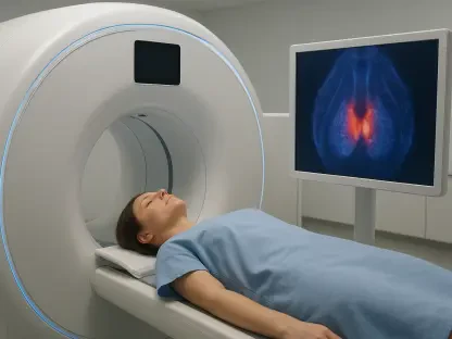

Recent clinical findings indicate that nearly half of patients with an initial negative scan undergo significant treatment modifications following a second imaging attempt. This discovery challenged the traditional reliance on a single diagnostic window, highlighting a critical blind spot in the management of biochemical recurrence. For many years, a negative result from a prostate-specific membrane antigen (PSMA) PET scan left both physicians and patients in a state of clinical limbo, where rising PSA levels suggested the return of cancer, yet imaging failed to locate the source. The transition toward a more iterative diagnostic process represented a fundamental shift in oncological strategy. Medical teams utilized the temporal progression of the disease to their advantage. This methodology recognized that the molecular expression of PSMA can fluctuate or intensify over time, making a secondary scan a vital reassessment that often revealed previously occult lesions in the pelvic region.
Enhanced Detection Strategies for Recurrent Disease
The Importance of Biometric Timing
The efficacy of repeat imaging is deeply intertwined with the biological behavior of the tumor, particularly regarding the specific threshold of prostate-specific antigen in the bloodstream. In 2026, the clinical community observed that waiting for a modest increase in these levels, rather than rushing to a different modality, often yielded the most actionable data. When the second scan was performed at a slightly higher PSA velocity, the sensitivity of tracers like Gallium-68 PSMA-11 or Pylarify increased exponentially. This strategic patience allowed oncologists to distinguish between a false negative and a true absence of disease. By analyzing the doubling time of the biomarkers, specialists could schedule the follow-up scan at a moment when the tumor’s metabolic activity was likely to surpass the detection limit of the scanner. This approach minimized the period of uncertainty, ensuring that salvage therapies were initiated at the optimal time, preventing the premature administration of systemic drugs.
Technical Resolution and Sensitivity
Technological advancements in the hardware used for these procedures further augmented the success of secondary imaging efforts. Modern PET/CT and PET/MRI systems available in 2026 utilized digital photon counters and advanced reconstruction algorithms that significantly improved the signal-to-noise ratio. These enhancements were particularly effective during a second look, as they allowed radiologists to visualize minute lymph nodes and small bone lesions that were once considered below the resolution threshold. The integration of artificial intelligence for image interpretation also played a role, as these systems could compare the first and second scans to identify subtle changes in uptake intensity that a human eye might overlook. This precision facilitated the use of stereotactic body radiation therapy (SBRT), allowing for the delivery of ablative doses to localized spots while sparing the surrounding healthy organs. The resulting data shifted the narrative from broad-spectrum treatment to a highly targeted, localized strike.
Clinical Implications for Personalized Therapy
Evolution of Diagnostic Standards
Integrating repeat PSMA PET scans into the standard diagnostic pathway redefined the categorization of recurrent prostate cancer. Previously, many cases with an initial negative scan were reflexively categorized as “non-visible” disease, often leading to generalized radiation of the prostate bed or lifelong hormone suppression. However, the high conversion rate of negative to positive findings upon re-imaging proved that a significant portion of these patients actually had oligometastatic disease. Identifying these specific sites allowed for a pivot in the therapeutic plan, moving away from palliative care and toward a curative intent. This shift necessitated a change in institutional protocols, where the failure of an initial scan to find a lesion was no longer seen as a diagnostic dead end. Instead, it became a baseline for future comparison. The ability to pinpoint a single involved node changed the prognosis for thousands of patients, offering them a chance at long-term remission that was previously deemed unlikely.
Strategic Implementation and Future Outcomes
The widespread adoption of this iterative imaging strategy successfully optimized the use of medical resources and improved patient survival rates. Healthcare providers emphasized the importance of standardized follow-up intervals, ensuring that no patient with a rising PSA was left without a secondary diagnostic path. It was recommended that medical centers invest in the latest digital PET technology to maximize the detection potential of every scan performed. Furthermore, the clinical workflow was adjusted to include multi-disciplinary reviews where radiologists and urologists collaboratively analyzed the timing of repeat scans based on individual patient kinetics. These actions ensured that diagnostic persistence remained a cornerstone of oncology, providing a clear roadmap for salvage interventions. By refusing to settle for the limitations of a single snapshot, the medical community established a more resilient framework for managing recurrence. This commitment to diagnostic thoroughness provided patients with the most accurate information.