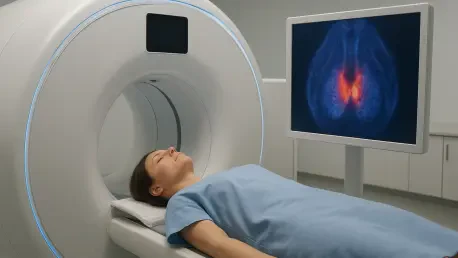

The diagnosis of metastatic medullary thyroid carcinoma is frequently hindered by the high physiological background activity of liver tissue in standard 18F-DOPA scans. This specific challenge is particularly problematic for patients with medullary thyroid carcinoma (MTC), a rare neuroendocrine malignancy that originates in the parafollicular C cells of the thyroid. While MTC accounts for a small fraction of all thyroid cancers, its management remains exceptionally complex because the primary curative approach is limited to surgical intervention. Once the disease spreads or recurs, localized treatment becomes a matter of precision, yet clinicians often find themselves in a diagnostic blind spot. Many patients present with rising levels of biochemical markers, such as calcitonin or carcinoembryonic antigen, while conventional imaging fails to pinpoint the exact location of the recurrent lesions. This creates a desperate need for more sensitive imaging modalities that can identify hidden metastatic sites to facilitate effective clinical planning.
The Biological Rationale: Targeting the Tumor Stroma
Traditional molecular imaging for neuroendocrine tumors typically focuses on the metabolic or secretory pathways of the malignant cells. For instance, the established clinical standard has been 18F-DOPA-PET, which tracks the amine precursor uptake and decarboxylation process within the cancer cells themselves. However, a significant paradigm shift is occurring with the introduction of tracers that target the fibroblast activation protein (FAP), an enzyme that is highly expressed in cancer-associated fibroblasts. These fibroblasts are key components of the tumor microenvironment, forming a supportive structural stroma that facilitates tumor growth and invasion. By shifting the diagnostic focus from the cancerous cells to the surrounding environment, FAP-targeted ligands like [68Ga]BED003 offer a unique way to visualize the malignancy. This method provides ultra-high affinity for the tumor stroma, potentially revealing the physical presence of recurrent disease in cases where traditional metabolic imaging might be inconclusive.
The recent development of [68Ga]BED003 represents a notable technological advancement in this field, utilizing a small-molecule ligand designed for high-precision binding. In contemporary research, this FAP-targeted tracer has been utilized as a problem-solving tool in clinically enriched cohorts where standard 18F-DOPA-PET scans were insufficient. By applying this technology to patients with suspected recurrence but negative standard scans, researchers are exploring how the tumor microenvironment can serve as a more reliable diagnostic target. This strategy is particularly useful for identifying the underlying cause of rising tumor markers when traditional imaging shows no visible abnormalities. The use of both PET/CT and PET/MRI systems in these evaluations has allowed for a comprehensive assessment of how these tracers behave in real-world clinical scenarios, providing a clearer understanding of their potential to redefine the current diagnostic pathways for managing persistent and metastatic medullary thyroid carcinoma.
Quantitative Advantages: Enhanced Detection Rates
Recent quantitative analyses have demonstrated a stark difference in the performance of FAP-targeted imaging compared to traditional methods. In comparative studies, [68Ga]BED003-PET identified a significantly higher percentage of lesions than 18F-DOPA-PET, with detection rates reaching nearly 97% in some cohorts compared to less than half for the established reference. This disparity is largely driven by the superior standardized uptake values (SUV) provided by the FAP-targeted tracer. The images produced by this new method are notably brighter and more distinct, allowing radiologists to assess the metabolic tumor volume with much greater precision. Such high intensity not only simplifies the identification of small lesions but also provides a more accurate representation of the total disease burden. This clarity is essential for surgeons who require an exact roadmap of the disease before proceeding with complex neck dissections or other localized interventions.
A critical factor in the success of FAP-PET is the dramatically improved tumor-to-background ratio (TBR) it offers across various tissues. In traditional imaging, physiological background noise in organs like the liver can often mask the presence of metastases, leading to false-negative results. The [68Ga]BED003 tracer effectively eliminates this issue by providing “cleaner” images with minimal tracer uptake in healthy liver, lung, and muscle tissues. This high contrast is particularly valuable in the liver, where the TBR for FAP-PET has been recorded as being significantly higher than that of traditional tracers. By providing a sharp distinction between malignant tissue and healthy organs, FAP-targeted imaging ensures that small metastatic sites are not overlooked due to the natural activity of the body. This robustness makes it a powerful asset for whole-body staging, offering a level of sensitivity that was previously unattainable with conventional metabolic tracers in the thyroid.
Site-Specific Superiority: Imaging Difficult Anatomical Regions
The clinical utility of FAP-PET is most evident when examining anatomical regions that are notoriously difficult to image using traditional methods. For instance, the liver and lungs often present significant hurdles due to high background activity or motion-related artifacts. In recent evaluations, [68Ga]BED003-PET successfully identified all liver metastases in study groups where traditional 18F-DOPA-PET was unable to detect a single one. This success is primarily attributed to the low physiological uptake of the FAP ligand in the liver, which allows for a high-contrast visualization of any stromal activity related to the tumor. Similarly, in the lungs, the FAP tracer has shown a superior ability to identify small nodules, overcoming challenges related to respiratory motion and partial-volume effects. This suggests that FAP-targeted imaging could become the preferred method for mapping out systemic spread in the abdomen and chest for thyroid cancer patients.
Beyond the major organs, FAP-PET has also proven to be highly effective in detecting bone metastases and lymph node involvement. All bone lesions identified in recent comparative studies were visualized using the FAP-targeted method, whereas traditional scans missed a substantial portion of these occurrences. This comprehensive sensitivity is vital for determining the extent of the disease and for identifying patients who might benefit from palliative radiation or other bone-targeted therapies. When it comes to lymph node mapping, the FAP tracer provides a detailed overview of regional spread, which is a critical component of surgical planning. While traditional methods are relatively effective at finding larger nodal involvement, the enhanced sensitivity of FAP-PET allows for the detection of smaller, more elusive nodes. This detailed mapping is essential for ensuring that surgical interventions are as thorough as possible, reducing the risk of further recurrence.
Addressing Diagnostic Pitfalls: Specificity and Complementary Use
While the high sensitivity of FAP-PET is a major advantage, it is important to acknowledge the potential for diagnostic pitfalls related to tracer specificity. FAP expression is not exclusive to malignancy; it can also occur in various inflammatory and fibrotic conditions. For example, tracer uptake has been observed in mediastinal lymph nodes caused by inflammation, such as anthracosis, which could be mistaken for cancer if the images are interpreted in isolation. This highlights the necessity of using FAP-PET as a complementary tool rather than a standalone diagnostic. Clinicians must interpret these scans alongside other cross-sectional imaging modalities like MRI or CT to correlate tracer uptake with anatomical findings. This multi-modal approach helps to differentiate between true malignancy and benign inflammatory processes, ensuring that patients are not subjected to unnecessary treatments based on false-positive results.
There are also instances where traditional tracers like 18F-DOPA-PET may provide unique insights that a FAP-only approach might miss. For example, traditional neuroendocrine tracers are capable of identifying secondary conditions like pheochromocytomas, which can sometimes occur alongside medullary thyroid carcinoma as part of a genetic syndrome. Furthermore, occasional lesions might show higher metabolic activity than stromal activity, leading to rare cases where 18F-DOPA-PET identifies a node that FAP-PET does not clearly visualize. These nuances suggest that the most effective diagnostic strategy involves a personalized selection of tracers based on the patient’s specific clinical profile. Rather than replacing established protocols, FAP-targeted imaging should be viewed as a high-powered specialized asset that adds a new dimension to the diagnostic toolkit, particularly in complex cases where rising tumor markers remain unexplained by current standard procedures.
Future Potential: Theranostics and Clinical Integration
The impressive tumor-to-background contrast of [68Ga]BED003 has sparked significant interest in the field of theranostics, a strategy that combines diagnostic imaging with targeted therapy. The concept involves using the same FAP-targeting molecule to deliver therapeutic radionuclides, such as Lutetium-177 or Actinium-225, directly to the tumor microenvironment. However, recent data indicated that while the tracer is excellent for visualization, many lesions did not meet the high intensity thresholds currently required for effective radioligand therapy. Specifically, the SUVmax levels in many cases remained below the benchmarks established for therapeutic intervention. This suggests that while [68Ga]BED003 is an outstanding diagnostic instrument, further optimization of the ligand or higher dosing strategies may be necessary to transform it into a viable vehicle for delivering localized radiation. Research in this area is ongoing to determine how FAP-targeted molecules can be best utilized for treatment.
Moving forward, the clinical integration of FAP-PET into standard thyroid cancer care will require more extensive validation. Current findings, while promising, were often based on small, specialized patient groups, which might not fully reflect the broader population of medullary thyroid carcinoma patients. Prospective, multicenter trials are essential to confirm these initial results and to establish clear clinical guidelines for the use of FAP-targeted imaging. These future studies should focus on how this technology impacts long-term patient outcomes and survival rates. Medical institutions began prioritizing the standardization of FAP-PET protocols to ensure consistency in reporting and interpretation across different clinical settings. The transition toward this technology was marked by a growing consensus that stromal targeting provides a vital second look for difficult cases. Ultimately, the integration of these advanced imaging techniques established a more precise and personalized approach to managing one of the most challenging forms of thyroid cancer.