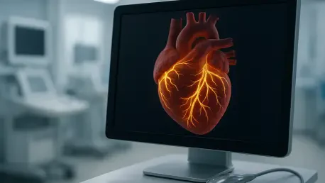

By leveraging standard ultrasound technology, Electromechanical Wave Imaging provides a noninvasive method for detecting risk factors associated with sudden cardiac arrest. For decades, the medical community has struggled to predict when manageable heart conditions like Mitral Valve Prolapse might escalate into lethal events. Mitral Valve Prolapse and its frequent companion, Mitral Regurgitation, involve the failure of the heart’s valve leaflets, causing blood to leak backward into the atrium rather than flowing forward. While millions of people live with these conditions, a small percentage faces an elevated risk of sudden cardiac death due to underlying electrical instability. Current diagnostic tools, including standard echocardiograms, often fail to detect these subtle warning signs because they focus on the heart’s anatomy rather than the intricate coordination between its electrical signals and muscle movements. This gap in diagnostic capability has left many patients in a state of uncertainty, waiting for symptoms that may arrive during a crisis.
Pioneering Cardiac Diagnostic Technology
Mapping: Mapping the Heart’s Electrical and Mechanical Coordination
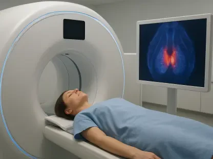
Elisa Konofagou and her research team at Columbia Engineering have introduced Electromechanical Wave Imaging, or EWI, to bridge the critical gap between structure and function. Unlike traditional ultrasound which merely visualizes blood flow and valve movement, EWI captures the propagation of the electrical wave as it moves through the myocardium. This technique utilizes high-frame-rate ultrasound to monitor the minute physical deformations that occur immediately after an electrical impulse triggers the muscle. By documenting these events at a millisecond level, the technology allows clinicians to see exactly how the heart’s internal wiring dictates its mechanical performance. This dual-layered perspective is essential for identifying the specific “short circuits” that lead to dangerous arrhythmias. Traditional imaging often isolates these components, but EWI integrates them into a single, comprehensive map that highlights the fundamental relationship between electricity and motion.
Precision: Visualizing Internal Wiring and Physical Pumping
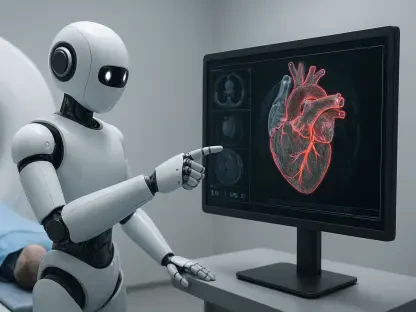
The sophistication of EWI lies in its ability to pinpoint the exact origin of contraction within the heart walls. In a healthy heart, the electrical impulse follows a highly organized path, ensuring that different chambers contract in a synchronized fashion to maximize efficiency. When valve diseases like Mitral Valve Prolapse are present, this timing can be disrupted, leading to localized mechanical stress that eventually causes electrical remodeling. EWI reveals these disruptions by color-coding the sequence of activation across the heart’s surface, making it easier for doctors to identify regions that are lagging or firing prematurely. This high-resolution mapping provides a level of detail that was previously only available through invasive catheter-based procedures or expensive, time-consuming MRI scans. By making this information accessible through standard equipment, the technology democratizes advanced cardiac care, allowing for more frequent monitoring and more precise intervention strategies.
Research Methodology and Patient Observations
Validation: Isolating Disease Indicators in Diverse Age Groups
To validate the effectiveness of Electromechanical Wave Imaging, researchers conducted a comprehensive study focusing on both pediatric and adult populations. By including children with Mitral Valve Prolapse, the team was able to study the disease in its purest form, unclouded by the secondary complications of aging, such as coronary artery disease or long-term hypertension. This approach allowed the scientists to determine if the electrical irregularities were a primary feature of the valve disorder itself or merely a result of cumulative wear on the heart. The results indicated that even in very young patients, EWI could detect significant delays in the electrical activation of the heart muscle. These findings suggest that the risk factors for sudden cardiac death may be present much earlier in life than previously suspected. Understanding these early-stage changes is vital for developing long-term management plans that can adapt as the patient grows and matures.
Stratification: Distinguishing Between Prolapse and Regurgitation
One of the most significant breakthroughs of the research was the ability to distinguish between the electrical impacts of Mitral Valve Prolapse and Mitral Regurgitation. While these conditions often occur together, they put different types of stress on the heart muscle. Patients with Mitral Valve Prolapse experience a mechanical tugging on the papillary muscles as the valve leaflets bulge backward. EWI demonstrated that this physical pulling leads to localized delays in electrical conduction. In contrast, Mitral Regurgitation involves a volume overload where blood leaks back into the left atrium, stretching the heart chamber over time. The study found that patients with significant regurgitation exhibited much longer recovery times for their electrical signals compared to those with prolapse alone. This distinction is crucial for risk stratification, as it suggests that the volume of blood leakage might be a more significant driver of electrical instability.
Clinical Implementation and Future Outlook
Standard: Enhancing Early Detection and Standardized Care
The ultimate goal for the medical community is to integrate Electromechanical Wave Imaging into the standard software packages of the ultrasound scanners already found in clinics and hospitals. Since EWI relies on the same hardware as traditional echocardiograms, the barrier to adoption is primarily software-based, making it a highly scalable solution for global healthcare systems. As this technology becomes more widely available, it will empower primary care cardiologists to perform advanced screenings during routine checkups. Early intervention is the key to preventing sudden cardiac death, and having a noninvasive, cost-effective tool to identify at-risk patients is a major step forward. Future updates to these systems will likely include automated analysis tools that use machine learning to flag abnormal patterns, further streamlining the diagnostic process and ensuring that life-saving technology is not restricted to specialized centers.
Evolution: Improving Long-Term Heart Health Management
Looking back at the progress made since the initial clinical trials, the medical field moved closer to a proactive model of heart health management. Researchers continued to expand their datasets, including more diverse patient populations to ensure the timing metrics were accurate across different ethnicities and lifestyles. The focus shifted toward creating standardized protocols that allowed doctors to compare EWI results over several years, tracking the slow progression of valve disease with unprecedented clarity. By 2026, the integration of these electromechanical markers into routine care successfully provided a clearer pathway for surgical timing and medication adjustments. Clinical teams utilized these insights to prevent the onset of lethal arrhythmias in patients who previously would have gone undetected. The transition from reactive treatment to proactive monitoring proved to be the most effective strategy for reducing the incidence of sudden cardiac death.