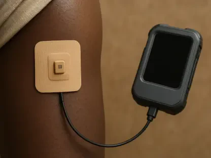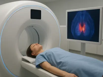

The interval between annual screenings is a critical vulnerability for many women, particularly those with dense breast tissue where aggressive tumors can manifest rapidly and without warning. Researchers at MIT have addressed this medical bottleneck by developing a wearable, portable 3D ultrasound scanner that shifts the paradigm from clinical dependence to proactive personal care. Under the leadership of Associate Professor Canan Dagdeviren, this technological breakthrough aims to bridge the screening gap that often leaves patients vulnerable to fast-growing interval cancers. By shrinking high-end diagnostic hardware into a handheld form factor, the team provides individuals with a means to monitor their health continuously. This democratization of clinical-grade imaging ensures that changes in tissue morphology are detected in their earliest stages, potentially saving lives by catching malignancies before they become symptomatic or spread to other parts of the body during the long wait for the next scheduled mammogram.
Overcoming Technical Barriers: Hardware and Software Engineering
The technical hurdles of condensing a large hospital-grade ultrasound machine into a portable device required a complete rethink of internal hardware components to maintain image quality. One significant challenge in miniaturization is managing electrical interference and stray mechanical vibrations that typically blur the resulting images and render them useless for diagnostic purposes. To solve this, the MIT engineers incorporated a specialized backing layer that effectively absorbs these unwanted signals, allowing the sensor array to function at higher power levels without sacrificing clarity. This advancement is crucial for penetrating deep into the breast tissue, reaching depths of nearly nine centimeters to visualize potential lesions that might be hidden by dense layers. By isolating the transducers from external noise, the device captures high-resolution data on microcalcifications, which are often the earliest precursors to cancerous growths. This level of precision was previously reserved for stationary machines costing hundreds of thousands of dollars.
Beyond the physical hardware, the system utilizes advanced beamforming software designed to navigate the heterogeneous nature of human breast tissue, which consists of varying ratios of fat and fibrous material. Sound waves travel at different speeds through these different tissues, causing standard ultrasound echoes to become smeared and lose detail as they travel back to the sensor. The MIT team developed a real-time algorithm that calculates the local speed of sound layer-by-layer, allowing the system to refocus the image dynamically based on the specific composition of the user’s body. This computational approach results in a 10% improvement in spatial resolution compared to conventional handheld probes, matching the performance metrics of full-sized clinical equipment. Such high-fidelity imaging is essential for distinguishing between harmless cysts and suspicious masses, providing the user with a reliable baseline for long-term health tracking. This synergy of robust hardware and intelligent software ensures that the portable device is not merely a gadget but a legitimate medical tool.
Empowering Patient Autonomy: Guided Technology for Home Use
Transitioning complex medical technology into the home environment necessitates a simplified interface that compensates for the user’s lack of professional training in sonography. The MIT researchers addressed this by creating the MyFUS guidance system, an innovative software layer that uses a standard computer webcam to track the ultrasound probe’s physical position in space. During an initial setup, the software records the specific coordinates and orientation used for a baseline scan, creating a digital roadmap for all subsequent sessions. When the user performs a follow-up check, the interface provides real-time visual prompts to help align the device perfectly with the previous scan site, ensuring that data is gathered from the exact same anatomical region every time. This focus on repeatability is what allows the device to detect subtle changes over several months with a high degree of confidence, reaching up to 90% accuracy in longitudinal tracking. By removing the guesswork associated with handheld scanning, the system empowers individuals to take an active role in their own diagnostic journey.
The efficacy of this guidance system was rigorously tested using volunteers who possessed no prior medical background or experience with ultrasound technology. These participants were tasked with finding microscopic targets hidden within breast models that mimicked the density and complexity of human tissue. The results were remarkable, as users successfully located 80% of the hidden targets, a success rate that far exceeded those using conventional handheld probes without the MyFUS tracking system. This high success rate is largely attributed to the device’s wide imaging block, which covers a larger surface area and makes it significantly harder for a user to accidentally skip over a small lesion or area of concern. The stability provided by the device’s form factor reduces the physical fatigue and hand tremors that often compromise the quality of scans performed by novices. This user-friendly interface represents a significant leap forward in home-based preventative care. Consequently, the technology serves as a reliable safety net, providing peace of mind for those at high risk of breast cancer.
Validating Clinical Utility: Future Directions in Healthcare
To ensure that the device met the stringent requirements of the medical community, the research team conducted extensive clinical validation in partnership with professional radiologists. These experts confirmed that the portable 3D scanner could accurately distinguish between various types of tissue features, including benign lumps, fluid-filled cysts, and medical implants, with the same level of nuance as hospital equipment. Safety was another primary concern, specifically regarding the thermal effects of prolonged ultrasound exposure on human skin and internal tissue. Testing proved that the device maintained temperatures well within established medical safety limits even after thirty minutes of continuous operation, making it safe for regular home use. These validation steps demonstrate that the device is not only technically capable but also practically viable for integration into the broader healthcare ecosystem. By proving its reliability in a controlled clinical setting, the researchers have paved the way for the technology to be adopted as a standard adjunct to traditional mammography for high-risk patients.
The successful development of this portable ultrasound platform established a foundation for future medical applications that extend far beyond breast cancer monitoring. By proving that high-resolution 3D imaging could be miniaturized and operated by non-professionals, the researchers opened new avenues for remote diagnostics in underserved regions. The next phase of this project focused on integrating the scanner with mobile technology, allowing users to sync their data directly with smartphones for immediate analysis by artificial intelligence or remote physicians. This capability allowed for the tracking of tumor shrinkage during chemotherapy treatments, providing doctors with real-time feedback on a patient’s response to specific drugs. Furthermore, the core technology was adapted to screen for other conditions, such as ovarian cancer or monitoring fetal development in areas where hospital access remained limited. Medical organizations recognized that adopting such decentralized tools empowered patients to become active participants in their recovery, fundamentally changing the landscape of preventative medicine.