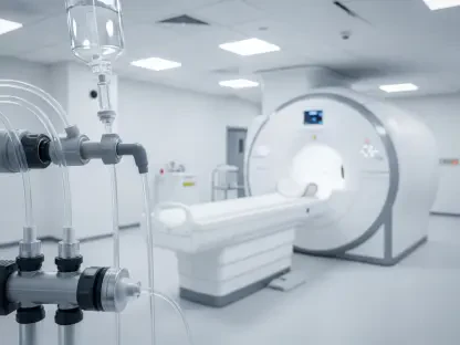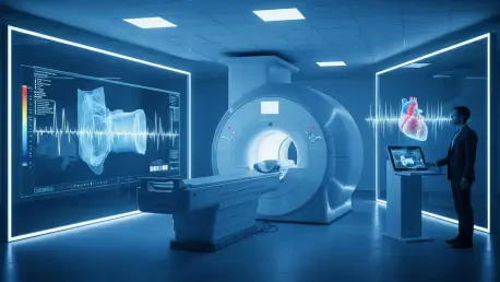

The traditional process of diagnosing complex heart conditions has long been hampered by the sheer volume of data produced during a single cardiac magnetic resonance imaging session. While these scans provide an unparalleled look at the heart’s internal mechanics, the burden of interpreting thousands of individual images often leads to significant delays in patient care and restricted access to specialized diagnostics. However, a recent breakthrough from Carnegie Mellon University and the Cleveland Clinic has introduced a system known as CMR-CLIP, which utilizes domain-specific foundation models to fundamentally change how this data is processed. By moving away from the rigid, labor-intensive labeling methods that have historically slowed artificial intelligence development, this new system leverages existing clinical narratives to achieve a more nuanced understanding of cardiac health. This shift represents a pivotal moment in medical technology, where the focus moves from simple pixel recognition to a deeper comprehension of the heart as a dynamic, living organ within a complex clinical context.
Bypassing the Manual Labeling Bottleneck
Learning Through Clinical Narratives
Standard approaches to training medical artificial intelligence have typically required thousands of hours of manual labor, as experts must meticulously label specific pixels or regions within an image to teach the software what to look for. This “data labeling” hurdle has long been a primary obstacle in the field of cardiac imaging, where the expertise required to identify subtle abnormalities is both expensive and rare. CMR-CLIP bypasses this bottleneck by utilizing the vast repositories of radiology reports already stored in hospital databases, specifically focusing on the “impression” section where clinicians summarize their findings in natural language. By training the model to align moving MRI sequences with these descriptive summaries, the researchers have enabled the system to learn the visual hallmarks of various pathologies through the collective wisdom of thousands of previous physician interpretations. This method effectively allows the AI to develop a medical vocabulary that mirrors human expertise.
The genius of this approach lies in its ability to associate complex visual patterns with professional medical terminology without the need for explicit, human-drawn boundaries on every frame of a scan. Instead of being told exactly which part of an image represents a scar or a valve defect, the model observes the relationship between the descriptive text of a report and the corresponding visual data in the video sequence. Over time, it learns that phrases such as “myocardial infarction” or “enlarged left ventricle” correspond to specific motion patterns and tissue densities. This training strategy not only accelerates the development process but also ensures that the AI remains grounded in real-world clinical practice. By tapping into the existing narrative history of cardiology, CMR-CLIP transforms static archives into a dynamic educational resource, providing a scalable solution that can be adapted to various medical institutions without the prohibitive costs of manual data curation or expert-led labeling.
Motion Analysis and Temporal Data
Unlike many previous iterations of diagnostic software that treated medical scans as a series of disconnected snapshots, CMR-CLIP treats cardiac MRI as a continuous, dynamic process. The heart is an organ defined by its motion, and a static image can only convey a fraction of the information necessary for a complete diagnosis. By processing time-resolved sequences, the system captures the intricate behavior of heart tissues and the fluid dynamics of blood flow throughout the entire cardiac cycle. This focus on temporal behavior allows the AI to mimic the actual workflow of a cardiologist, who must observe how the heart contracts and relaxes to assess pumping efficiency and structural integrity. The model’s ability to analyze these “videos” rather than just pictures provides a more comprehensive view of cardiac health, ensuring that subtle functional abnormalities are not missed during the screening process. This temporal awareness is critical for identifying conditions that only manifest during specific phases of the heartbeat.
Furthermore, the integration of motion analysis allows the system to evaluate the heart’s functional capacity with a level of precision that was previously difficult to achieve through automated means. By understanding how different regions of the heart muscle move in coordination, CMR-CLIP can pinpoint localized areas of dysfunction that might indicate early-stage disease or recovery from previous trauma. This depth of analysis is essential for managing patients with chronic conditions where tracking minute changes in cardiac output is vital for adjusting treatment plans. The system’s architectural design prioritizes the temporal relationship between frames, ensuring that every millisecond of the scan contributes to the final diagnostic impression. This approach not only enhances the accuracy of the findings but also provides a more robust framework for evaluating complex cases where static images might be ambiguous. By aligning the AI’s observational capabilities with the physics of a beating heart, the technology provides a more reliable foundation for clinical decision-making.
Establishing Performance and Clinical Reach
Superior Accuracy and Scalability
In rigorous comparative testing, the performance of CMR-CLIP has demonstrated the clear advantages of using domain-specific models over general-purpose artificial intelligence for specialized medical tasks. The system outperformed broader AI frameworks by more than 35%, achieving accuracy levels as high as 99% in specific diagnostic categories. One of the most significant findings was the system’s “zero-shot” learning capability, which allows it to identify rare or complex cardiac conditions it was never explicitly trained to recognize. By matching new images against descriptive prompts derived from its narrative training, the AI can extrapolate its knowledge to assist with unique patient presentations. This ability to generalize beyond its initial training set is a major leap forward, suggesting that the model can remain useful even as new medical insights and terminology emerge within the field of cardiology, effectively future-proofing the diagnostic tool.
To ensure that the system’s effectiveness was not merely a result of being tailored to a single institution’s data, the research team validated CMR-CLIP against diverse, independent datasets from locations as varied as France and Florida. The model maintained a high level of robustness across different patient demographics and varied imaging equipment, proving that it can handle the technical inconsistencies often found in global healthcare environments. This scalability is crucial for widespread adoption, as it suggests that the technology can be deployed in community hospitals and smaller clinics that may not have the same resources as major academic medical centers. By providing a consistent and high-quality “second opinion,” the system helps to democratize access to advanced cardiac diagnostics. This ensures that a patient’s geographic location does not dictate the quality of the interpretation they receive, ultimately leading to more equitable healthcare outcomes across different populations and regions.
Future Roadmap and Diagnostic Integration
Looking toward the immediate horizon, the research team is focused on expanding the capabilities of CMR-CLIP to include a wider array of imaging sequences and diagnostic functions. Current plans involve integrating perfusion imaging to monitor blood flow in real-time and parametric mapping to characterize the cellular composition of heart tissue. These additions will allow the system to provide even deeper insights into conditions like myocarditis or rare infiltrative diseases, where tissue characterization is just as important as structural analysis. The ultimate goal is to create a fully integrated “reader assistant” that can automatically generate preliminary reports and highlight areas of concern for the attending physician. By handling the more routine aspects of scan interpretation, the AI allows human experts to focus their attention on the most complex and critical aspects of patient care, significantly reducing the administrative and diagnostic burden currently facing the cardiology community.
As the technology continues to mature, its integration into clinical decision support systems will likely transform the way cardiologists interact with historical patient data. The system’s natural language processing capabilities could enable clinicians to search through thousands of past cases using simple descriptive phrases, allowing them to find “digital twins” or similar clinical presentations for comparison. This capability is particularly valuable when dealing with rare diseases where a single physician may only see a handful of cases in their entire career. By providing instant access to a vast library of analyzed scans and outcomes, CMR-CLIP facilitates a more informed and data-driven approach to individual patient management. To maximize the impact of these advancements, healthcare organizations should begin evaluating their data infrastructure to support seamless AI integration. Investing in interoperable systems and standardized reporting will be essential steps for clinics looking to adopt these transformative tools and improve the speed and accuracy of cardiac diagnostics.