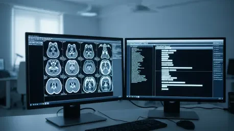

The rapid intersection of advanced computer vision and large language models has fundamentally altered the landscape of neuro-oncology, providing a glimpse into a future where diagnostic delays are largely eliminated. While traditional methods for identifying genetic markers in brain tumors have historically relied on surgical intervention, a new era of medical technology has arrived with the development of Glio-LLaMA-Vision. This multimodal artificial intelligence system represents a significant shift in how medical professionals approach adult-type diffuse glioma, a particularly aggressive form of brain cancer. By analyzing standard magnetic resonance imaging scans, this system can now predict the presence of the isocitrate dehydrogenase gene mutation with remarkable precision. This capability effectively bridges the gap between initial imaging and molecular diagnosis, which typically requires weeks of laboratory analysis. The integration of these complex capabilities into a single workflow signifies a profound evolution in clinical practice, moving beyond simple image recognition toward a more holistic understanding of tumor biology.
Advancing Neuro-Oncology Through Multimodal Intelligence
Eliminating the Need for Invasive Procedures
The primary challenge in managing adult-type diffuse glioma has always been the necessity of obtaining physical tissue samples to determine the specific genetic makeup of a tumor. Biopsies are not only invasive and carry risks of infection or neurological deficit, but they are also costly and time-consuming for both the patient and the healthcare facility. The Glio-LLaMA-Vision system disrupts this traditional paradigm by utilizing non-invasive MRI data to extract molecular insights that were previously hidden from the naked eye. By identifying subtle patterns in the imaging that correspond to the IDH mutation, the AI provides a level of diagnostic depth that rivals traditional pathology. This transition from physical sampling to digital analysis allows for a safer patient experience while maintaining the high standards of accuracy required for oncology. The ability to bypass the surgical risks associated with tissue extraction marks a significant milestone in the journey toward truly non-invasive cancer diagnostics.
Beyond the immediate safety benefits, the speed at which this AI operates allows for the rapid initiation of personalized treatment plans. In many clinical settings, waiting for genomic sequencing can delay the start of chemotherapy or targeted radiation for several weeks, during which time the tumor may continue to progress. With the implementation of this vision-language model, clinicians can receive a high-probability molecular profile almost immediately after the imaging session is complete. This acceleration is particularly critical for glioma patients, where survival rates are heavily influenced by how quickly the correct therapeutic strategy is applied. The system achieved an impressive Area Under the Curve score between 0.85 and 0.95, demonstrating that its diagnostic reliability is consistent across diverse patient populations. Such high accuracy levels ensure that the shift toward AI-driven diagnostics does not compromise the quality of care but instead enhances the precision of the initial medical assessment.
Automating the Radiology Workflow
One of the most innovative aspects of this technological breakthrough is its ability to generate comprehensive radiology reports that are ready for clinical use. Traditionally, radiologists must spend a significant portion of their day meticulously documenting every observation from an MRI scan, a process that is prone to fatigue and human error. Glio-LLaMA-Vision automates this descriptive task by translating visual data into structured medical narratives that follow professional standards. Approximately 90% of the reports generated by the system were deemed suitable for clinical application by veteran medical specialists, highlighting the sophistication of its language processing capabilities. This automation does not merely save time; it ensures that every report maintains a high level of consistency and detail, regardless of the workload volume at a particular hospital. By handling the heavy lifting of documentation, the AI allows radiologists to focus their expertise on the most complex aspects of patient care.
The creation of these automated reports involves a complex synchronization between image interpretation and medical linguistics. The model was trained on vast repositories of biomedical literature from sources like PubMed Central, enabling it to understand the nuanced terminology and syntax required in neuro-oncology. This deep training allows the AI to describe tumor margins, enhancement patterns, and edema with the same level of nuance as a human professional. Furthermore, the system employs a preprocessing step to normalize sentence structures, which prevents the variability that often plagues automated text generation. This ensures that the final output is not just a collection of keywords, but a coherent and professional document that integrates seamlessly into a patient’s electronic health record. The result is a more efficient diagnostic pipeline where the communication between the imaging department and the oncology team is faster and more standardized than ever before.
Integrating Genetic Data into Clinical Decision Making
Refining Diagnostic Accuracy with Specialized Training
The success of Glio-LLaMA-Vision is rooted in its specialized fine-tuning process, which utilizes actual brain tumor MRI images paired with standardized clinical reports. Unlike general-purpose AI models, this system is specifically designed to navigate the intricacies of the central nervous system and the unique presentation of malignant gliomas. By training on thousands of cases, the model has learned to distinguish between various tumor grades and molecular subtypes that might appear identical to an untrained observer. This specialized training allows the AI to pick up on “radiomic” features—data points within the image that are too subtle for human vision to detect. These features often correlate strongly with the genetic status of the tumor, providing a digital signature of the IDH mutation. This level of specialization is what allows the model to achieve such high AUC scores, proving that a targeted approach to AI development is essential for solving complex medical problems.
Furthermore, the integration of vision and language within a single model allows for a more contextual understanding of the patient’s condition. The AI does not look at the image in a vacuum; it understands how specific visual features should be described and what they imply for the patient’s prognosis. This dual-modality approach mimics the cognitive process of a radiologist, who must synthesize visual information into a logical medical conclusion. By mimicking this human-like reasoning, the AI provides a more reliable output that clinicians can trust when making critical decisions about surgery or medication. The research, supported by the Ministry of Science and ICT, highlights a growing consensus that multimodal systems are the future of digital medicine. These systems provide a bridge between raw data and actionable knowledge, ensuring that the wealth of information contained within a single MRI scan is fully utilized to benefit the patient’s long-term health outcomes.
Streamlining Hospital Operations and Patient Care
The implementation of Glio-LLaMA-Vision has broader implications for the operational efficiency of healthcare systems, particularly in high-volume cancer centers. By reducing the time required for both molecular diagnosis and report generation, hospitals can process a greater number of patients without increasing the burden on their existing staff. This efficiency is vital in an era where healthcare resources are often stretched thin and the demand for specialized imaging continues to rise. The system acts as a force multiplier for the radiology department, providing a high-quality “first draft” of every report and a preliminary genetic assessment that the doctor can then verify. This collaborative approach between human and machine reduces the cognitive load on medical professionals, potentially lowering the rates of burnout among specialists. As a result, the entire diagnostic pathway becomes more resilient and better equipped to handle the complexities of modern neuro-oncological care.
Moreover, the clinical utility of this AI extends to the patient’s understanding of their own condition. Automated reports that are clear, standardized, and timely can help in explaining the diagnosis to the patient and their family, facilitating more informed discussions about the next steps in treatment. When the molecular status of a tumor is known early on, the conversation can shift immediately to the most effective therapies rather than waiting in a state of uncertainty. This transparency is a crucial element of patient-centered care, as it empowers individuals to be active participants in their treatment journey. The transition toward these intelligent systems represents a commitment to modernizing healthcare through technology that prioritizes both precision and speed. By streamlining the path from the initial scan to the final treatment plan, the medical community is setting a new standard for how technology can improve the human experience in the face of a challenging diagnosis.
The emergence of Glio-LLaMA-Vision as a viable clinical tool suggests that the next phase of neuro-oncology will be defined by the seamless integration of molecular biology and artificial intelligence. Medical institutions should begin exploring the infrastructure requirements for hosting multimodal AI systems, ensuring that local data privacy standards are met while maximizing the computational power necessary for real-time analysis. Future research should focus on expanding this model to include a wider variety of genetic markers and tumor types, potentially creating a universal diagnostic assistant for all forms of brain cancer. Clinicians must also undergo specialized training to effectively interpret and validate AI-generated reports, maintaining the essential human oversight that ensures patient safety. As these technologies continue to mature from 2026 toward 2030, the goal will be to establish a global standard where non-invasive genetic profiling becomes the primary method of initial diagnosis. By adopting these tools, the healthcare industry can ensure that every patient receives a personalized, data-driven treatment strategy from the moment they first enter an imaging suite.