
In the high-stakes environment of a modern trauma center, where minutes determine patient outcomes and clinical teams are pushed to their limits, the arrival of more sophisticated imaging technology represents a fundamental shift in emergency medical operations. Medical professionals frequently encounter complex scenarios, ranging from severe vehicular accidents to sudden neurological events, necessitating tools that offer both extreme speed and uncompromising clarity. The introduction of the Rembra CT aims to address these specific pressures by integrating robust hardware with sophisticated artificial intelligence to transform how critical care is delivered. By prioritizing the intersection of speed and precision, the platform simplifies the most demanding workflows found in acute care settings. This development allows radiologists and surgeons to receive actionable data almost instantaneously, effectively removing the traditional bottlenecks that often occur during the triage process.
Global Launch: Regulatory Standards and Physical Foundations
The international medical community first witnessed the capabilities of this new imaging system during its official unveiling in March 2026 at the European Congress of Radiology in Vienna. This event served as a major platform for demonstrating how the technology could be integrated into diverse healthcare systems across the globe. Following the successful exhibition, the system rapidly secured 510(k) clearance from the US Food and Drug Administration, alongside the CE Mark for distribution within the European Union. These regulatory milestones are significant, as they validate the scanner’s adherence to the most stringent safety and performance standards in the industry. By obtaining these certifications so close to its launch, the platform entered the market ready for immediate deployment in major medical centers. This swift transition from debut to approval highlights a commitment to providing hospitals with verified tools that are prepared to handle the rigors of modern diagnostics and acute care.
The physical architecture of the system was engineered to solve the most persistent challenges in high-volume trauma centers, beginning with a massive 85cm bore that stands as the largest in its technical category. This extra-wide opening is essential for accommodating bariatric patients and providing medical staff with the necessary room to perform interventional procedures without the usual physical constraints of a CT gantry. Complementing this spacious design is a heavy-duty patient table that supports a scan range exceeding seven feet, which allows for comprehensive head-to-toe imaging in a single pass. These mechanical features collectively enable a high-throughput environment, where a single facility can reliably conduct up to 270 examinations in a single twenty-four-hour period. By prioritizing physical versatility and hardware durability, the platform ensures that hospital staff can treat a diverse patient population with consistent speed and anatomical coverage.
Diagnostic Precision: Advanced Detection and Workflow Automation
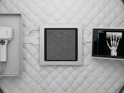
Achieving superior diagnostic clarity relies on the integration of the NanoPanel Precise 4cm XD detector, which utilizes advanced AI to maximize image resolution while significantly reducing the required radiation dose. This technology allows clinicians to capture minute anatomical details as small as 0.25mm, which is particularly vital for examining complex structures like the inner ear or subtle bone fractures. To maintain this high level of detail when scanning larger patients, an integrated anti-scatter grid works to preserve image contrast and clarity, ensuring that every scan meets the same rigorous quality standards. By focusing on micro-level precision, the detector provides radiologists with the fine-grained data necessary for making confident diagnoses in acute care settings where every detail matters. This hardware-driven accuracy is further enhanced by AI algorithms that optimize the signal-to-noise ratio, providing a crystal-clear window into the patient’s condition.
Clinical productivity is further maximized through specialized AI tools such as the Trauma Suite and the Precise Brain application, which automate repetitive tasks such as labeling vertebrae and identifying rib fractures. These automated features are designed to reduce the cognitive burden on medical staff, allowing them to shift their focus from manual data entry to higher-level clinical decision-making. Complementing this software is a set of patient-side controls that allow technologists to set up and manage scans directly at the gantry, saving valuable time and improving the overall patient experience. Furthermore, the inclusion of high-speed “zero-click” reconstruction ensures that images are processed at a rate of 106 per second and delivered instantly to the hospital’s digital archives. By streamlining the entire imaging sequence from setup to storage, the platform helps clinical teams manage the heavy patient loads typical of metropolitan emergency departments.
Strategic Integration: Operational Resilience and Long-Term Value
Looking toward the long-term sustainability of medical infrastructure, the scanner is built with a projected service life of 20 years, making it a cornerstone investment for any healthcare facility. The engineering team designed the hardware to function reliably under extreme conditions, including high-altitude environments that often pose challenges for sensitive electronic components. This robustness is matched by a high degree of versatility, as the system can seamlessly transition between standard diagnostic radiology and complex radiation therapy planning within the same department. This multi-functional capability allows hospital administrators to consolidate their imaging resources without sacrificing the specialized performance required for different clinical paths. By providing a platform that is both durable and adaptable, the technology ensures that healthcare providers can maintain a high standard of patient care while realizing a significant return on their capital expenditure.
Healthcare administrators and clinical directors evaluated the long-term benefits of adopting this automated imaging technology to address persistent gaps in emergency department efficiency. The decision to integrate these systems followed a detailed assessment of how AI-enhanced diagnostics could stabilize patient throughput during periods of peak demand. Strategic leaders focused on training radiological teams to leverage the automated labeling features, ensuring that the transition from legacy equipment was both smooth and immediately beneficial to patient safety. By investing in these resilient platforms, organizations prioritized the creation of a diagnostic infrastructure that was capable of adapting to future healthcare trends and rising patient volumes. This proactive approach allowed medical centers to establish a more streamlined workflow that reduced staff burnout and enhanced the overall quality of care delivered to the community. Ultimately, the implementation process served as a model for how advanced technology can be successfully utilized to modernize frontline medical services.