
A/Prof Zuber’s vision for veterinary medicine involves expanding PET-CT services nationally to make metabolic imaging the standard first step in cancer investigation. The establishment of PET Theranostics in Sydney represents a fundamental turning point for animal healthcare across the continent. By integrating advanced technology typically reserved for high-end human oncology, this facility bridges a longstanding gap in veterinary diagnostic capabilities. For years, clinicians often struggled to differentiate between active disease and scar tissue using conventional methods, but the arrival of specialized nuclear medicine has changed the equation. The center, founded by industry veterans with decades of expertise, offers a diagnostic depth that allows for much more personalized treatment plans. This evolution toward precision medicine ensures that companion animals receive care that is as sophisticated as the treatments available to their human counterparts, ultimately raising the bar for the entire profession in the current landscape.
Technical Synergy: Merging Anatomical and Metabolic Insights
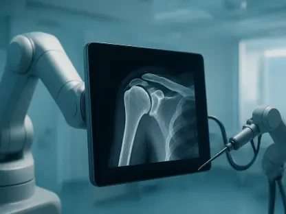
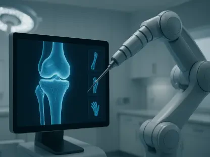
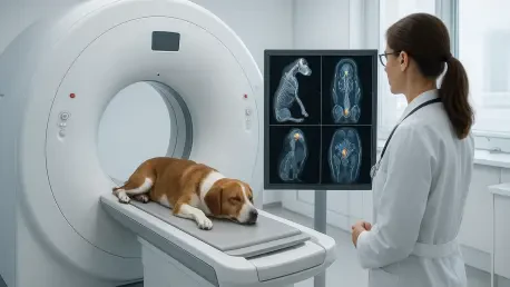
The technological backbone of this new era in veterinary care is the PET-CT scanner, a sophisticated dual-gantry system that integrates two distinct types of imaging into a single clinical session. This complex machinery operates by utilizing specific radioisotopes produced in a cyclotron, which are injected into the animal before the procedure begins. These isotopes have a notably short half-life, ensuring they are processed quickly by the body while providing the necessary signal for the detectors. The CT component of the scanner functions as the anatomical foundation, producing high-resolution images that define the physical structures of the patient, including bones, organs, and various soft tissues. Meanwhile, the PET component focuses on detecting the radiation emitted by the isotopes, which naturally gravitate toward areas of high metabolic activity. This dual-layered approach provides a much more comprehensive view of the patient’s internal health than any basic scanner.
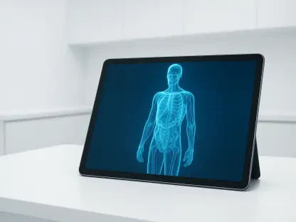
The true diagnostic power of this technology emerges from the digital fusion of the two separate data captures into a single, cohesive map of the animal’s body. By overlaying metabolic data directly onto the high-definition anatomical structural map, veterinarians can pinpoint the exact location of cellular abnormalities with unprecedented accuracy. This is particularly vital when dealing with malignant tumors, which are characterized by rapid and aggressive cellular growth. Traditional imaging techniques, such as standard X-rays or basic ultrasounds, often struggle to distinguish between a benign growth and a truly active malignancy. However, the PET-CT scan provides a clear metabolic signature that reveals the functional state of the tissue in question. This level of precision allows for far more accurate surgical planning and biopsy targeting, as clinicians can focus their efforts on the most active areas of the disease rather than relying on structural appearances alone.
Clinical Scope: Transforming Oncology and Internal Diagnostics
Oncology serves as the most immediate and impactful application for PET-CT in the current veterinary environment, as the technology excels at identifying the high metabolic rates typical of cancer. Beyond the initial diagnosis, the system has proven to be an invaluable tool for monitoring how well a patient is responding to ongoing therapy. For example, if a dog has undergone extensive radiation treatment for a brain tumor, a follow-up scan can determine if any remaining mass is still viable cancer or simply inert scar tissue. A low uptake of radioisotopes in a previously treated area is a strong indicator that the disease has been successfully eradicated, providing owners with much-needed clarity. This “theranostics” approach—which combines therapeutics with diagnostics—uses specialized isotopes to not only identify the primary tumor but also to track and deliver targeted radiation to metastatic sites that might be scattered throughout the body.
While oncology remains a primary focus, the clinical applications for PET-CT are expanding into other complex medical fields, including neurology and cardiology. In the neurological sphere, the technology is helping specialists diagnose conditions that parallel human neurodegenerative disorders, such as Alzheimer’s disease, by mapping changes in brain metabolism over time. In cardiology, the scanner is used to assess the viability of heart muscle, providing critical information for managing chronic cardiac disease in aging pets. Furthermore, the ability to identify occult inflammation makes the technology a powerful tool for veterinary sports medicine. It allows clinicians to detect subtle muscular issues or hidden inflammatory processes in highly active working animals or competitive athletes. By adapting techniques from human sports science, veterinarians are now able to provide a level of diagnostic insight that was previously impossible for most pet owners.
Safety Standards and Economic Future of Animal Care
Addressing public concerns regarding radiation exposure is a critical component of integrating nuclear medicine into mainstream veterinary practice. Despite common misconceptions, the PET-CT procedure is remarkably safe for the patient, with a radiation profile that is often lower than the dose received during a standard, full-body diagnostic CT scan alone. This safety was ensured by a collaborative framework that included radiation physicists from the human medical sector and specialized technicians from Ecuador for the initial system setup. To ensure that every scan is interpreted with the highest level of expertise, the clinic established an international network with radiologists from the University of Colorado who possess extensive experience in animal PET imaging. This partnership allowed for a secondary level of review that remains crucial when dealing with complex cases, overcoming the technical hurdles that previously hindered the adoption of this technology.
The move toward advanced nuclear medicine was supported by evolving economic trends, as pet owners increasingly prioritized high-level care despite the significant costs involved. While a PET-CT study typically ranged between $5,000 and $6,000, the proliferation of comprehensive pet insurance made these services accessible to a wider demographic of the population. This financial shift allowed the Sydney facility to attract “destination clients” who traveled from across the country to seek definitive answers for their pets’ health. Consequently, the team successfully demonstrated that there was a robust market for high-end diagnostics when paired with clear clinical outcomes. As the flagship clinic stabilized its operations, the vision for a national network of metabolic imaging centers moved closer to reality. Industry groups even explored industrial applications for the technology, such as studying livestock metabolism to improve overall agricultural health standards.