
The transition from intensity-based to volume-based metabolic assessment represents a significant shift in how oncologists might predict early treatment failure. While traditional staging systems rely heavily on tumor size and lymph node status, they often overlook the metabolic intricacies that signal how a tumor will behave post-surgery. Current medical strategies aim to provide curative treatment for early-stage breast cancer, yet a notable percentage of patients still experience early relapse. This discrepancy highlights a critical need for predictive tools that go beyond anatomical measurements. Recent findings published in Frontiers in Oncology suggest that preoperative [¹⁸F]FDG PET/CT imaging could serve as a powerful crystal ball. By analyzing the metabolic activity of tumors before the first incision is made, clinicians might better identify individuals at high risk for recurrence or metastasis within 24 months, allowing for more personalized and aggressive intervention strategies from the very beginning.
Methodology and Patient Selection Criteria
The research team focused on a cohort of 106 women diagnosed with clinical stage I or II breast cancer who were treated at Beijing Shijitan Hospital. To ensure the integrity of the metabolic data, the researchers implemented strict exclusion criteria, removing any patients who had already undergone neoadjuvant chemotherapy, radiotherapy, or targeted treatments. This step was essential because such therapies can significantly alter glucose metabolism within a tumor, potentially masking its true aggressive nature and skewing the PET/CT readings. By selecting patients who had not yet received systemic treatment, the study was able to capture a raw biological snapshot of the cancer. This methodology allowed for a clear baseline, ensuring that the subsequent imaging parameters were a direct reflection of the tumor’s inherent characteristics rather than a response to medical intervention. The follow-up period spanned two years, providing enough time to monitor early progression.
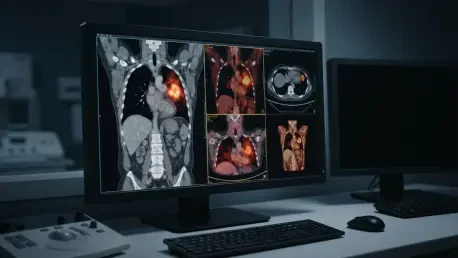
Standardization of the imaging protocol was paramount to achieving reproducible results across the study population. Every participant followed a strict preparation regimen that included a six-to-eight-hour fast and a controlled rest period after receiving an intravenous dose of [¹⁸F]FDG. Using a high-resolution Discovery 710 PET/CT scanner, the medical team performed three-dimensional acquisitions from the head to the mid-thigh. This comprehensive scanning approach combined low-dose CT for anatomical localization with PET data to visualize metabolic activity. Advanced reconstruction algorithms were applied to ensure that the quantitative data—such as the metabolic tumor volume—was as precise as possible. This technical rigor transformed a standard imaging procedure into a sophisticated diagnostic tool capable of quantifying the metabolic burden of the disease. The resulting data provided a robust foundation for comparing the imaging profiles of those who remained healthy against those who faced early recurrence.
Understanding Metabolic Imaging Parameters
Beyond the traditional visual inspection of scans, the researchers utilized three distinct quantitative metrics to evaluate the tumors: standardized uptake value (SUV), metabolic tumor volume (MTV), and total lesion glycolysis (TLG). While SUVmax has long been the standard for measuring the highest point of metabolic activity, it only represents a single pixel of the tumor’s intensity. In contrast, MTV provides a measurement of the actual physical volume of the tumor that is metabolically active, offering a better sense of the overall tumor burden. TLG further refines this by multiplying the MTV by the average metabolic intensity, effectively calculating the total glucose consumption across the entire lesion. These volume-based metrics are increasingly seen as more representative of the total biological weight of the cancer. Instead of just looking at how bright a tumor appears, these parameters quantify the scale of its metabolic machinery, providing a more holistic view of the threat.
The evaluation process extended beyond the primary breast lesion to include the ipsilateral axillary lymph nodes and the whole-body metabolic burden. Even in cases where the axillary nodes did not show obvious signs of disease on initial scans, the investigators employed a standardized region-of-interest method to document baseline activity. This level of detail is crucial because microscopic spread to the lymph nodes is a primary driver of recurrence in early-stage breast cancer. By capturing the metabolic activity of both the primary tumor and the surrounding lymphatic system, the study aimed to see if a systemic footprint was present even before clinical symptoms emerged. This multi-site analysis allowed for the calculation of whole-body MTV and TLG, which represents the cumulative metabolic activity of all detectable cancerous sites. This approach suggests that the risk of recurrence is not just about the primary mass, but the total amount of active disease present throughout the patient’s body.
Identifying Patterns in Cancer Recurrence
The outcomes of the 24-month follow-up revealed a significant divide in the patient population, with nearly 46% of the participants experiencing some form of recurrence or metastasis. The events recorded were diverse and often aggressive, including regional lymph node spread, bone involvement, and lung metastasis. When these outcomes were mapped back to the preoperative PET/CT data, clear patterns emerged that separated the recurrence group from those who remained cancer-free. Specifically, patients who faced a relapse had shown significantly higher primary tumor metabolic volumes and total glycolysis during their initial scans. For example, the average PT-MTV in the recurrence group was measurably larger than in the non-recurrence group. These findings suggest that a tumor’s metabolic footprint is a harbinger of its future behavior. The data clearly indicated that tumors with higher energy requirements and larger active volumes were much more likely to breach the initial surgical defenses.
One of the most insightful findings was that the peak intensity of the primary tumor, or SUVmax, did not significantly differ between the two groups. This highlights a limitation of traditional PET/CT interpretation, which often prioritizes the brightest spot in a lesion. The fact that volume-based measures like MTV and TLG were predictive while intensity-based measures were not suggests that the sheer quantity of active cancer cells is a better indicator of risk than the metabolic rate of a single cell. This shift in perspective is vital for oncologists who rely on imaging to guide treatment intensity. If a tumor is small but extremely active throughout its entire volume, it may pose a greater risk than a larger tumor with only localized activity. These results reinforce the idea that the total metabolic burden is a smoking gun for early treatment failure, providing a more reliable signal for clinicians to watch for potential relapses before they occur.
Biological Markers and Risk Factors
The study also explored the relationship between imaging parameters and the underlying biological markers that drive cancer growth. Patients in the recurrence group were significantly more likely to exhibit lymphovascular invasion, a condition where cancer cells are caught in the act of migrating into the circulatory or lymphatic systems. Furthermore, these patients often had higher levels of Ki-67 expression, which is a key indicator of rapid cellular proliferation. This high rate of cell division requires a substantial amount of energy, which explains the high glucose uptake seen on the PET/CT scans. There was a clear synergy between the pathological evidence of an aggressive tumor and the metabolic data captured through imaging. This correlation suggests that PET/CT is essentially providing a non-invasive look at the molecular aggressiveness of the disease, allowing doctors to estimate a tumor’s growth potential and invasive capability without waiting for extensive biopsy results.
Molecular subtypes also played a major role in the observed outcomes, with the recurrence group showing a higher prevalence of HER2-positive and triple-negative breast cancers. These specific subtypes are notoriously difficult to treat because they often lack the hormone receptors that respond to standard endocrine therapies. Interestingly, the study found that tumors with lower estrogen and progesterone receptor positivity tended to have higher metabolic volumes and glycolysis scores. This biological reality underscores why metabolic imaging is such a potent tool; it reflects the aggressive reality of these receptor-poor tumors that are more prone to early relapse. By identifying these high-risk biological profiles through preoperative imaging, medical teams can better prepare for the possibility of a more stubborn disease. The integration of imaging and pathology provides a multi-layered understanding of the patient’s risk, ensuring that the most dangerous tumors are flagged for closer monitoring and more intensive care.
Statistical Modeling and Predictive Accuracy
To confirm that these PET/CT measures were truly independent predictors of recurrence, the researchers employed multivariable logistic regression models. These models were designed to control for other clinical factors like tumor stage and lymphovascular invasion. The results were striking: for every one-unit increase in certain metabolic parameters, the odds of the cancer returning increased significantly. Specifically, an increase in the metabolic tumor volume of the primary lesion was associated with a much higher risk of recurrence. In some instances, a significant rise in metabolic volume was shown to quadruple the likelihood of treatment failure. This statistical evidence moves the conversation from general observation to concrete risk assessment. It proves that the metabolic data provides unique information that cannot be gathered from tumor size or lymph node status alone, making it a critical addition to the prognostic toolkit used by modern oncologists in 2026.
While individual metrics like MTV and TLG were useful on their own, the researchers found that combining multiple metabolic parameters into a single predictive model offered the highest level of accuracy. Using Receiver Operating Characteristic analysis, the study showed that a combined model involving primary tumor metrics and lymph node activity achieved a significantly higher area under the curve than any single measure. This high level of discriminatory power suggests that a metabolic profile is far more effective than a single data point. By integrating various snapshots of the cancer’s energy use, clinicians can achieve a much clearer picture of the patient’s prognosis. This multifaceted approach reflects the complexity of breast cancer itself, acknowledging that no single factor determines the outcome. Instead, it is the cumulative effect of tumor volume, intensity, and nodal involvement that provides the most accurate forecast of whether a patient will remain in remission or face a new challenge.
Limitations and Future Research Needs
Despite the significant progress made in this study, there are several limitations that must be addressed before these techniques can be fully integrated into daily clinical practice. The research was retrospective and conducted at a single medical center with a relatively small sample size of 106 patients. This means that the findings, while compelling, need to be validated through larger, prospective trials involving diverse populations from different geographic regions. Without such validation, it is difficult to determine if these metabolic thresholds would hold true in a broader clinical context. Additionally, the study did not have an external validation cohort, which is the gold standard for proving the reliability of a predictive model. Future research will need to focus on establishing standardized cut-off values for MTV and TLG that can be used across different imaging centers, ensuring that a high-risk score in one hospital means the same thing in another facility.
Another important consideration is the 24-month follow-up period, which focuses exclusively on early recurrences. While identifying early treatment failure is vital, many types of breast cancer—particularly hormone-receptor-positive cases—can recur many years after the initial diagnosis. Long-term studies extending well beyond the current timeframe will be necessary to see if these preoperative metabolic measures can also predict late-stage relapses. Furthermore, the researchers noted that variations in postoperative adjuvant treatments, such as different chemotherapy regimens or the duration of hormone therapy, were not fully accounted for in the analysis. These treatments can significantly alter the trajectory of the disease, potentially masking the predictive power of the initial imaging. Future studies should aim to integrate treatment variables into their models to see how metabolic risk interacts with modern systemic therapies, providing a truly comprehensive view of patient outcomes.
Practical Implications for Patient Care
The study successfully demonstrated that preoperative metabolic assessments provided a window into the biological behavior of breast cancer that traditional staging often missed. By identifying patients with a high total metabolic burden, medical teams were able to flag individuals who required more vigilant surveillance or potentially more aggressive adjuvant therapy. This shift toward personalized risk stratification represents a major advancement in clinical oncology, moving away from a one-size-fits-all approach. The evidence suggested that using PET/CT as a prognostic tool allowed for a more nuanced understanding of which tumors were likely to resist standard surgical and medical interventions. Ultimately, the data supported the idea that the volume of metabolic activity was a critical factor in determining the likelihood of a successful long-term outcome, providing a new metric for success that went beyond the removal of the visible primary tumor mass.
Looking forward, the integration of these metabolic parameters into standard preoperative workups was identified as a key step for improving survival rates. Clinical practitioners were encouraged to look beyond the peak intensity of a scan and instead focus on the total metabolic volume as a primary indicator of risk. Actionable steps included the development of automated software tools that could quickly calculate MTV and TLG, making these metrics more accessible to busy oncology departments. Furthermore, hospitals were advised to begin incorporating these imaging biomarkers into multidisciplinary tumor board discussions to better tailor post-operative care. By treating high metabolic scores as a call to action for increased monitoring or enhanced therapeutic protocols, the medical community took a significant step toward reducing the burden of early recurrence. This proactive approach paved the way for more refined treatment pathways that specifically addressed the unique metabolic profile of every individual patient.