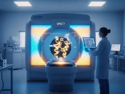

The landscape of vascular medicine underwent a radical shift when clinical researchers introduced a molecular imaging technique capable of mapping every thrombus in the human body simultaneously. For decades, medical professionals relied on fragmented snapshots provided by disparate imaging modalities, often missing the broader context of a patient’s circulatory health. However, the emergence of a specialized positron emission tomography (PET) radiotracer has redefined the diagnostic standard by moving beyond simple structural visualization to detect active clot formation at its cellular source. This breakthrough, which was recently recognized as the “Image of the Year” at a prominent nuclear medicine symposium, offers a comprehensive view that was previously considered unattainable in a single clinical session. By focusing on the biochemical signatures of thrombosis, healthcare providers can now identify silent risks before they escalate into catastrophic events such as strokes or embolisms. This shift from physical to molecular detection marks a new era in preventative care.
Overcoming the Limitations: Why Current Diagnostics Often Fail
Current standard procedures for identifying deep vein thrombosis typically involve venous ultrasonography, a method that relies heavily on the physical compression of veins and the observation of blood flow patterns. While ultrasound remains a valuable tool due to its accessibility and lack of ionizing radiation, it suffers from significant limitations that can compromise patient safety in critical scenarios. The effectiveness of this technique is largely dependent on the expertise of the individual operator and the specific physical characteristics of the patient, leading to variability in results across different clinical settings. Furthermore, standard ultrasound is frequently restricted to specific segments of the limbs, meaning that a negative result in one area does not necessarily rule out the presence of a dangerous clot elsewhere in the body. This piecemeal approach often requires multiple scans and prolonged observation periods, delaying the initiation of essential anticoagulant therapies.
The diagnostic gap is particularly evident when considering the difficulty of detecting clots located in the calves or the complex pelvic region where ultrasound waves struggle to provide clear imagery. In many cases, patients presenting with vague symptoms may have extensive thrombotic activity that remains invisible to conventional technology until a life-threatening pulmonary embolism occurs. CT scans, while more comprehensive, often utilize contrast agents that can be taxing on the kidneys and still fail to distinguish between old, stable clots and new, active ones that pose the greatest risk. This lack of specificity means that clinicians must sometimes rely on clinical intuition or serial testing to make high-stakes decisions about surgical interventions or long-term medication. The inability to visualize the entire vascular system in a single, high-resolution pass has long been a significant hurdle in the management of complex cases involving multiple comorbidities or systemic inflammation.
Molecular Precision: The Impact of the 18F-GP1 Radiotracer
To address these diagnostic shortfalls, the development of the 18F-GP1 radiotracer has introduced a level of precision that targets the physiological essence of a blood clot rather than its shadow. This tracer is designed to bind specifically to activated platelets, which are the primary building blocks of a fresh thrombus, allowing the PET scanner to highlight areas of active clotting with intense clarity. Unlike traditional imaging that looks for the obstruction of a vessel, the 18F-GP1 PET scan identifies the molecular activity within the clot itself, regardless of its size or location. This capability enables physicians to see the full extent of a patient’s thrombotic burden in one comprehensive sweep, covering everything from the cranial vessels down to the extremities. Because the tracer ignores inactive or old clots, it provides a real-time assessment of the current threat level, ensuring that medical interventions are directed toward the most dangerous and unstable blockages currently in formation.
Clinical validation studies demonstrated that this molecular approach achieved a sensitivity of 95 percent and a specificity of 92 percent, confirming its reliability as a primary diagnostic tool for complex vascular conditions. The implementation of this technology allowed medical teams to identify clots in the heart valves, spine, and other unconventional sites that were routinely missed by localized ultrasound examinations. Looking ahead from 2026 to 2030, the focus shifted toward integrating this whole-body imaging into standard protocols for embolic stroke workups and high-risk cardiovascular screenings. Researchers recommended that future implementations prioritize the miniaturization of tracer production to make this high-tier diagnostic capability accessible to regional medical centers. By establishing a centralized database for molecular thrombotic profiles, the medical community paved the way for highly personalized treatment plans that optimized patient outcomes while minimizing risks.