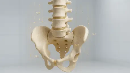

Mathematical filters like LASSO regularization ensure that AI models focus only on the most predictive markers of cancer while discarding irrelevant data points. This methodological precision is essential when addressing sacral tumors, which represent a significant hurdle in modern orthopedic oncology due to their deep-seated location and the intricate anatomy of the pelvic-spinal junction. Because these lesions are statistically infrequent, clinicians often struggle to differentiate between benign growths and aggressive malignancies based on symptoms alone, which frequently mimic common conditions like sciatica. Standard clinical presentations, such as persistent lower back pain or radicular discomfort, are often identical regardless of the tumor’s biological nature. This diagnostic ambiguity poses a significant risk, frequently leading to the invasive over-treatment of harmless masses or the dangerous under-treatment of life-threatening cancers. To resolve this, researchers at Peking University People’s Hospital have developed an approach using artificial intelligence to analyze non-contrast computed tomography scans.
Advanced Radiomic Methodology
The Digital Shift: Data-Centric Oncology
The shift toward data-centric diagnostic models has fundamentally altered how orthopedic oncologists approach the evaluation of the sacrum. In 2026, the traditional reliance on visual interpretation is being augmented by high-throughput computing that can process thousands of data points simultaneously. This transition is not merely a change in toolsets but a paradigm shift in how medical images are perceived—moving from static pictures to multidimensional datasets. The methodology developed at Peking University People’s Hospital leverages this evolution by focusing on the radiomic signature of tissue, which encompasses texture, intensity, and spatial relationships that the human eye cannot consistently quantify. By implementing a rigorous algorithmic pipeline, the research team has established a framework that minimizes subjective bias in tumor assessment. This advanced methodology serves as the foundation for the habitat-based analysis that follows, ensuring that the resulting diagnostic models are grounded in objective, reproducible data.
Exploring the Tumor Ecosystem: Habitat Partitioning
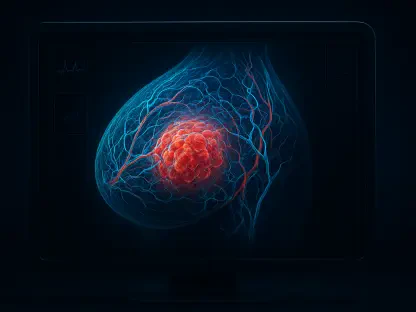
The core of this research is built upon the understanding of tumor heterogeneity, which posits that a tumor is not a uniform mass but a complex ecosystem of diverse sub-regions. Traditional imaging methods often treat a sacral lesion as a homogenous entity, averaging its characteristics and potentially missing the aggressive localized signals of malignancy. However, the use of habitat radiomics allows for the identification of specific zones within a tumor that exhibit unique biological behaviors. These sub-regions may indicate areas of rapid cell proliferation, oxygen deprivation, or even early-stage necrosis, all of which are critical indicators of how a tumor will likely progress. By acknowledging that different parts of a single tumor can behave differently, the AI model provides a far more nuanced assessment than conventional radiological reports. This internal diversity is a hallmark of cancerous growth, and capturing it mathematically is essential for accurate differentiation between benign and malignant sacral tumors.
Partitioning Techniques: Internal Density Analysis
Using a machine learning technique known as K-means clustering, the researchers successfully partitioned each sacral tumor into three distinct spatial habitats based on density variations. These partitions are determined by Hounsfield units, which measure the radiodensity of tissues in a standard computed tomography scan. The AI uses these values to create a digital map of the tumor’s internal architecture, highlighting clusters of high-density tissue and comparing them to more porous or cystic areas. This spatial mapping is supported by the Calinski-Harabasz criterion, ensuring that the divided habitats are statistically significant and biologically relevant. By focusing on these specific internal habitats rather than the entire lesion at once, the model can identify chaotic organizational patterns that are typical of malignant tumors. This method effectively transforms a standard scan into a detailed topographical map of the tumor’s biological landscape, providing clinicians with a level of insight that was previously available only through invasive tissue sampling.
Feature Selection: Streamlining Predictive Markers
To ensure the accuracy of the diagnostic model, the research team harvested thousands of quantitative features from the imaging data, including texture, shape, and wavelet-transformed intensities. However, processing such a vast amount of information carries the risk of overfitting, where a model becomes too specialized to its training set and loses its ability to predict new cases accurately. To mitigate this, the team employed LASSO regularization, which acts as a sophisticated filter to streamline the dataset. This mathematical process identifies and retains only the most predictive markers of cancer while discarding redundant or irrelevant data points that might skew the results. By narrowing down the features to a high-impact core, the researchers created a streamlined and robust algorithm capable of high-level performance in diverse clinical environments. This meticulous selection process is what allows the AI to distinguish the subtle noise of benign tissue from the definitive patterns of malignancy, ensuring that the resulting diagnosis is both reliable.
Algorithmic Performance: The Neural Network Model
After evaluating several machine learning architectures, the researchers determined that a Multi-Layer Perceptron neural network offered the most robust performance for classifying sacral tumors. This deep learning model is designed to recognize intricate, non-linear relationships between the extracted radiomic features and the final pathological diagnosis. The MLP’s ability to process complex interactions within the data allowed it to achieve an Area Under the Curve of 0.877 when focusing on tumor habitats. This significantly outperformed more traditional, single-entity models, proving that the spatial arrangement of a tumor’s internal sub-regions is far more diagnostically valuable than its total average density. The neural network also demonstrated that the 3-millimeter peri-tumoral shell provided a more critical diagnostic battleground than the 6-millimeter shell, identifying the exact zone where the tumor’s interaction with host bone is most indicative of malignancy. These findings underscore the importance of precision in both spatial analysis.
Clinical Application and Outcomes
Integrated Diagnostics: The Ensemble Nomogram
Translating complex algorithmic outputs into actionable clinical insights is the final and most critical step in this diagnostic evolution. While the technical performance of a neural network is impressive in a laboratory setting, its true value is measured by its impact on patient care and surgical outcomes. In 2026, the integration of AI into the clinical workflow at major hospitals has shown that data-driven models can bridge the gap between radiology and the operating room. The results of the Peking University study highlight how fused diagnostic tools—those that combine digital imaging features with established clinical variables—offer a more complete picture of a patient’s condition. This transition from raw data extraction to clinical application allows surgeons to move forward with a level of confidence that was previously unattainable. By evaluating the real-world utility of these models through rigorous statistical testing, the researchers have demonstrated that AI is an enhancer of expertise.
Decision Support: Real-World Clinical Utility
The integration of various data streams culminated in the creation of an ensemble nomogram, which combined the patient’s age with peri-tumoral data and internal habitat signatures. This fused model achieved an impressive Area Under the Curve of 0.892, a level of precision that represents a major advancement over traditional diagnostic methods. To put this in perspective, models based solely on clinical symptoms like patient age and the presence of pain achieved an AUC of only 0.619, which is only slightly better than a random guess. The ensemble approach succeeds because it recognizes that neither imaging data nor clinical history is sufficient on its own to provide a definitive diagnosis in the complex sacral region. By merging these different perspectives into a single, cohesive tool, the nomogram offers a comprehensive risk assessment that can be easily interpreted by surgical teams. This high degree of accuracy allows for more effective triage of patients, ensuring that those with malignant signatures are prioritized.
Patient Safety: The Accessibility of NCCT Scans
To confirm the real-world utility of the ensemble nomogram, the research team utilized Decision Curve Analysis to evaluate its net benefit in a clinical setting. This assessment is vital because a model can be statistically accurate without being practically useful for making difficult medical choices. The analysis revealed that the nomogram provided a high net benefit across a broad range of probability thresholds, meaning it successfully helped clinicians avoid unnecessary surgeries for benign tumors while ensuring that malignant ones were not overlooked. This calibration between predicted probabilities and actual pathological outcomes is essential for the tool’s adoption in surgical planning. By providing a clear, evidence-based probability of malignancy, the AI-driven model acts as a reliable decision-support system that reduces the psychological and physical burden on both patients and surgeons. This level of validation ensures that the technology moves beyond a research curiosity and becomes a practical instrument for improving clinical care.
Surgical Evolution: The Future of Virtual Biopsies
A primary advantage of this AI-driven diagnostic framework is its specialized focus on non-contrast computed tomography scans, which are widely available and carry fewer risks for the patient. Many advanced imaging protocols in 2026 require the use of contrast agents to enhance visual detail, but these agents can lead to allergic reactions or exacerbate existing kidney problems. By demonstrating that high-level diagnostic accuracy can be achieved using standard NCCT scans, the researchers have made sophisticated oncology tools more accessible to a global population. This approach is particularly beneficial for patients who have contraindications for contrast-enhanced imaging or for facilities that lack the specialized equipment required for more complex scans. Furthermore, because NCCT is a standard part of the initial workup for back pain, this AI model can be integrated into existing workflows without requiring additional procedures. This efficiency reduces the time patients must wait for a definitive diagnosis.
Technological Impact: Personalized Treatment Pathways
The development of the habitat radiomics model at Peking University People’s Hospital provided a transformative solution for the complex management of sacral tumors throughout 2026. By utilizing non-invasive imaging to reach a diagnostic certainty of nearly 90 percent, the technology allowed for a significant shift in surgical philosophy toward nerve-preserving strategies. These actionable insights ensured that patients with benign lesions avoided the life-altering complications of aggressive resections, such as the loss of bladder or bowel control. The integration of this AI tool into the clinical decision-making process established a new standard for personalized orthopedic oncology, where treatment was dictated by the specific biological reality of each tumor. Future efforts focused on expanding these datasets to include rarer tumor subtypes and validating the model across international centers to ensure global applicability. Ultimately, the success of this virtual biopsy approach demonstrated that the future of oncology resided in medical imaging.