
The rapid evolution of oncological care has reached a critical juncture where the sheer volume of diagnostic imaging often outpaces the capacity of human specialists to interpret complex longitudinal data with absolute precision. For years, clinicians relied on manual measurements and subjective visual assessments to track the progression of tumors, a method that is notoriously prone to inter-observer variability and significant delays in treatment adjustments. Today, the introduction of an AI-native oncology platform across select medical centers in the United States signals a profound shift toward automated, data-driven diagnostic frameworks that integrate seamlessly with clinical workflows. By leveraging advanced foundation models trained on massive datasets, this technology aims to provide instantaneous, standardized analysis of radiological scans, ensuring that every patient receives a level of scrutiny that was previously impossible. This deployment represents more than just a software update; it is a foundational change in how cancer centers approach the intersection of imaging and patient management.
Standardizing the Landscape: Advancements in Radiological Assessment
The Architecture of Innovation: Modern Foundation Models in Medicine
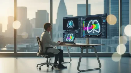
At the core of this technological leap lies a sophisticated foundation model specifically engineered to understand the nuances of human anatomy and the subtle patterns of oncological pathology. Unlike traditional AI tools that require specific prompts or narrow parameters to function, this AI-native platform operates with an inherent understanding of spatial relationships within the body, allowing it to detect anomalies across multiple organ systems simultaneously. This architectural shift means that radiologists no longer need to toggle between various disparate software packages to analyze a single patient’s case. Instead, the platform provides a unified interface where deep learning algorithms automatically segment tissues, identify suspicious lesions, and compare them against historical imaging data. By utilizing these multi-modal capabilities, the system reduces the cognitive load on physicians, permitting them to focus on the high-level interpretation of results rather than the tedious manual labor of measurement and documentation, thereby enhancing the overall quality of care.
The initial deployment of this platform at major academic medical centers across the United States has already provided valuable insights into its practical utility within high-volume clinical settings. By integrating these AI-native tools into the daily operations of departments at institutions like Massachusetts General Hospital, researchers are observing a significant reduction in the time needed to review complex oncology cases. These sites serve as a testing ground for the platform’s ability to handle diverse patient populations and varying imaging protocols, ensuring that the models remain robust and accurate across different hospital environments. The success of these early implementations is a testament to the collaborative effort between data scientists and clinical practitioners who worked to refine the user interface and ensure the output met the high standards of oncological practice. As more hospitals adopt this technology, the collective data generated will further improve the underlying models, creating a feedback loop that continually enhances the diagnostic precision available to every oncologist in the network.
Accuracy and Precision: Streamlining Clinical Decision Support
One of the most persistent challenges in modern radiology is the inherent variability in how different specialists interpret the same set of images, particularly when measuring small changes in tumor size or density. This inter-observer inconsistency can lead to discrepancies in staging and treatment planning, potentially impacting patient outcomes if a progression is missed or incorrectly identified. The platform addresses this issue by providing a standardized, objective baseline for every measurement, utilizing advanced segmentation algorithms that consistently apply the same criteria across all scans. By automating the application of standardized criteria like RECIST, the system ensures that every assessment is reproducible and verifiable, regardless of the individual radiologist’s experience level. This consistency is vital for clinical trials and routine care alike, as it provides a reliable metric for evaluating the efficacy of specific therapies over time. Reducing this margin of error allows oncology teams to make more confident decisions, knowing that the data guiding their strategy is both accurate and uniformly processed.
Beyond standardization, the platform significantly enhances diagnostic accuracy by identifying subtle radiological markers that might be overlooked by the human eye during a standard review. For instance, in the early stages of lung or liver cancer, lesions can be extremely faint or obscured by surrounding anatomical structures, making them difficult to detect without the aid of high-resolution computer vision. The AI-native system scans every pixel of the image with a high degree of sensitivity, flagging suspicious areas for the radiologist to investigate in more detail. This proactive approach acts as a powerful safety net, catching potential issues early when they are most treatable and reducing the likelihood of missed diagnoses. Furthermore, the platform’s ability to analyze longitudinal changes over several years allows it to spot slow-growing pathologies that might appear stable in the short term but show a clear trend over a longer horizon. By augmenting human perception with these sophisticated analytical capabilities, the technology sets a new benchmark for what can be achieved in modern oncological diagnostics.
Strategic Integration: Operational Impact and Clinical Efficacy
Seamless Connectivity: Bridging the Gap between Data and Delivery
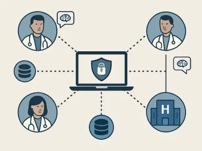
Integrating AI technology into the complex ecosystem of a modern hospital requires more than just clinical accuracy; it demands seamless interoperability with existing imaging and electronic health record systems. This platform is designed to sit directly within the radiologist’s native workflow, pushing and pulling data from the Picture Archiving and Communication System without requiring a separate login or manual data entry. This level of integration is crucial for maintaining high throughput in busy clinics, as any friction in the technology stack can lead to delays and user frustration. By automatically pre-populating reports with quantitative data and standardized findings, the platform allows radiologists to complete their reviews more quickly while maintaining a high level of detail. This efficiency gain is not merely about speed; it is about reclaiming time that can be redirected toward patient consultation and multidisciplinary tumor boards. When the administrative and technical burdens of imaging are minimized, the entire healthcare team can operate more effectively, leading to faster turnaround times for results.
The scalability of an AI-native oncology platform is another significant advantage, as it allows smaller community hospitals to access the same high-level diagnostic tools as large academic centers. Historically, the most advanced imaging analysis was restricted to institutions with the budget and expertise to maintain complex in-house software, but cloud-native AI solutions have democratized this access. By providing a scalable infrastructure that can be deployed rapidly across multiple sites, the technology ensures that patients in rural or underserved areas benefit from the same precision diagnostics as those in major metropolitan hubs. This democratization of technology is a vital step toward reducing health disparities in oncology care, as early and accurate diagnosis is one of the most significant factors in long-term survival. Moreover, the cost-effectiveness of an automated system becomes apparent as hospitals reduce the need for repeat scans and minimize the resources spent on manual data reconciliation. As these platforms become more widespread, the overall efficiency of the national healthcare system improves, creating a sustainable model for delivering high-quality cancer care.
Lessons in Adoption: Actionable Strategies for Institutional Implementation
As oncology moves toward more personalized treatment strategies, the ability to predict how an individual patient will respond to a specific therapy has become increasingly valuable. The AI-native platform contributes to this goal by identifying imaging phenotypes that are associated with positive or negative responses to immunotherapy and targeted treatments. By analyzing thousands of data points within a single scan, the system can uncover subtle patterns in tumor morphology and vascularity that serve as early indicators of therapeutic success or failure. This predictive capability allows oncologists to pivot their strategy sooner if a treatment is not working, sparing patients from unnecessary side effects and focusing resources on more effective alternatives. In the coming years, starting from 2026 and moving through the decade, the integration of genomic data with these advanced imaging biomarkers will likely lead to even more precise predictions. This multi-layered approach to patient analysis ensures that every therapeutic decision is backed by a robust combination of visual and biological evidence, moving us closer to the ideal of truly individualized medicine.
The successful implementation of these AI-native systems at initial clinical sites established a clear framework for the broader adoption of automated diagnostics in oncology. Medical directors at participating hospitals found that prioritizing a culture of technological literacy was essential for ensuring that staff members could effectively leverage the platform’s capabilities. Clinicians conducted thorough internal audits to compare AI outputs with traditional methods, which helped build the necessary confidence to incorporate these tools into standard practice. Furthermore, the technical teams ensured that data security protocols remained a top priority during the integration process, protecting sensitive patient information while maintaining high system performance. These institutions demonstrated that a phased approach, starting with specific cancer types before expanding to more complex cases, allowed for a smoother transition and better resource allocation. By following these established best practices, other healthcare providers successfully mitigated the challenges of modernization. The lessons learned from these early deployments created a sustainable path forward for the entire industry, emphasizing that the combination of human expertise and advanced technology is the most effective way to improve patient outcomes.