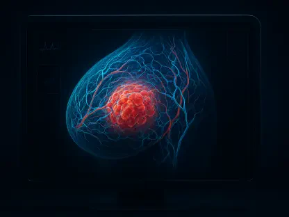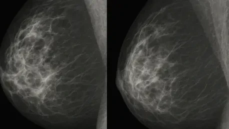

Heatmaps generated by explainability tools ensure that a model is looking at actual lesions rather than clinical markers or background artifacts. This development marks a pivotal shift in how the medical community views artificial intelligence in 2026, where the goal has moved beyond simple predictive accuracy to meaningful diagnostic transparency. Breast cancer remains a global health priority, with early detection consistently serving as the most significant variable in determining patient outcomes and long-term survival rates. While mammography has provided the backbone of screening for decades, the process remains fraught with challenges, as radiologists must distinguish subtle malignancies from dense breast tissue while navigating high caseloads. Recent research conducted at Selcuk University by Elham Tahsin Yasin, Ilkay Cinar, and Murat Koklu has demonstrated that sophisticated deep learning models can significantly mitigate these human limitations, reaching detection accuracies of up to 98 percent through optimized networks.
Strategic Methodology: Transfer Learning in Specialized Medical AI
To achieve such high precision without an infinite supply of medical data, researchers utilized a technique called transfer learning, which has become the standard for specialized medical imaging. This involves taking AI models already trained on millions of general images and repurposing them for complex oncology tasks. By leveraging the model’s existing ability to recognize basic visual features like edges, gradients, and textures, the system can quickly adapt to the specific patterns of malignancy in mammograms. This approach is essential in contemporary healthcare, where high-quality, expert-labeled data is often scarce due to strict privacy laws and the high cost of manual annotation. Transfer learning allows these networks to start with a foundation of visual literacy, which drastically reduces the amount of specialized training data required to reach professional diagnostic levels. This methodology effectively bridges the gap between general computer vision and the high-stakes requirements of clinical cancer detection.
Precision Benchmarking: Evaluating Convolutional Neural Network Architectures
The core objective of this specific research was to conduct an “explainability-aware” benchmarking of several prominent convolutional neural network architectures. The models selected for evaluation included DenseNet201, InceptionResNetV2, MobileNetV2, NASNetLarge, and Xception, each representing a different approach to processing visual information. By comparing these diverse structures, the researchers could identify which architectural features are most conducive to spotting the minute calcifications and structural distortions indicative of early-stage breast cancer. The evaluation focused not just on the final accuracy score but on how efficiently each model could learn from the available radiographic samples. This benchmarking process is critical for the industry, as it provides a clear roadmap for which AI structures should be prioritized for integration into hospital workstations. The goal was to find a balance between high-level sensitivity to cancerous cells and the specificity required to avoid unnecessary and stressful false positives for the patients.
Performance Optimization: The Impact of Sophisticated Data Augmentation
A critical element in reaching the 98 percent accuracy threshold was the use of data augmentation to expand the training set from 745 to nearly 10,000 images. Initially, the researchers worked with a relatively small dataset, which is a common bottleneck in medical AI development because it can lead to overfitting, where the model simply memorizes the training images. By applying transformations such as rotations, horizontal flips, and vertical reflections, the team forced the AI to become more robust and less sensitive to the specific orientation of a lesion. This ensured that the model learned the actual characteristics of malignancy rather than just the specific positioning of the tumors in the original photos. The results of this expansion were undeniable, as every architecture tested showed significant improvement in its diagnostic performance after the augmentation was implemented. This highlights the reality that in medical computer vision, the quality and quantity of data are just as important as the code itself.
Diagnostic Accuracy: Analyzing DenseNet and Architecture Convergence
The impact of data expansion was most visible in the performance of DenseNet201, which emerged as the leading architecture with a top accuracy of 98.06 percent when trained on the augmented data. DenseNet’s success is largely attributed to its dense connectivity design, where each layer receives feature maps from all previous layers, facilitating a more thorough reuse of visual information. However, the study also revealed a fascinating trend toward convergence; as the volume of training data increased, the performance gap between different AI architectures began to narrow significantly. This suggests that modern neural networks have reached a level of maturity where several different designs can achieve near-perfect binary classification if they are fed sufficient information. For the medical community, this means that the choice of specific software may eventually become secondary to the establishment of massive, high-quality data repositories that can be used to refine any authorized diagnostic system.
Global Health Implications: Efficiency in Resource-Limited Settings
Beyond the peak accuracy scores, the success of more lightweight models like MobileNetV2 carries profound implications for global healthcare equity. MobileNetV2 was designed for computational efficiency, yet it achieved nearly the same accuracy as its much heavier and more resource-intensive counterparts, such as NASNetLarge. This finding suggests that high-accuracy diagnostic tools could eventually be deployed in small clinics or resource-limited areas using standard hospital hardware rather than expensive server clusters. By reducing the computational cost of cancer detection, this technology democratizes access to high-level screening, ensuring that patients in remote locations can benefit from the same diagnostic precision as those in major metropolitan medical centers. This shift toward efficient, mobile-ready AI is a key step in reducing the global burden of breast cancer, as it allows for the implementation of screening programs in regions that historically lacked the specialized personnel or the budget for advanced imaging infrastructure.
Explainable Intelligence: Visual Activation Mapping for Clinical Trust
For artificial intelligence to be truly useful in a professional hospital setting, it cannot function as a “black box” where doctors are expected to trust a percentage score blindly. The researchers addressed this fundamental issue by using explainability-aware frameworks like Grad-CAM, which produce heatmaps that highlight the specific regions of an image that influenced the model’s decision. This transparency allows a radiologist to verify that the AI is actually focusing on a suspicious lesion rather than a random artifact or a clinical marker in the background. By providing this visual evidence, the AI transitions from a mysterious algorithm to a collaborative partner that assists the human expert in making a final diagnosis. This building of clinical trust is essential for the wide-scale adoption of AI, as it preserves the role of the physician while providing them with a high-speed, high-precision second opinion that can catch subtle details that might be missed during a long and tiring shift.
Statistical Rigor: Validating Models Through Cross-Validation Techniques
The study utilized stratified k-fold cross-validation to ensure that the 98 percent accuracy rate was a consistent result and not the product of a favorable data split. This rigorous statistical method involves dividing the data into multiple subsets and training the model several times, each time using a different subset for testing. By achieving high performance across all folds, the researchers proved that their models were highly generalizable and capable of handling new, unseen mammograms with the same level of precision. While these results are a landmark achievement, moving into the next phase of clinical adoption required even broader validation across diverse patient populations. Real-world screening involves varied breast densities, different types of imaging equipment, and a wide array of non-cancerous abnormalities that can mimic tumors. Ensuring that the AI remains accurate across these variables is the current focus of ongoing clinical trials, as the industry moves toward standardized, evidence-based integration of deep learning.
Strategic Implementation: Enhancing Survival Through Human-Machine Synergy
The integration of these high-accuracy models acted as a transformative force in the clinical landscape, where AI transitioned from a theoretical tool to a reliable second reader in radiology departments. This implementation primarily focused on triaging routine screenings, which allowed human experts to dedicate more of their cognitive resources to complex or ambiguous cases. By providing a consistent baseline of 98 percent accuracy, the technology helped to standardize care across various hospital settings, regardless of the individual experience level of the attending physician. Furthermore, the systematic use of explainability-aware frameworks ensured that every diagnostic output was backed by visual evidence that a clinician could verify in real time. This collaborative approach proved essential for reducing false negatives, which historically posed the greatest risk during the early stages of oncological development when intervention is most effective for patient survival.
Future Standards: Lessons Learned from High-Precision AI Development
Researchers and medical practitioners ultimately found that the primary value of these neural networks lay in their ability to maintain peak performance without the fatigue that often compromises human perception during long shifts. As high-quality, open-source datasets became more prevalent, the technical barriers to deploying these systems in resource-limited environments were dismantled, enabling a global improvement in screening standards. The success of this methodology encouraged healthcare administrators to prioritize the development of more robust data-sharing agreements, which facilitated the training of even more generalized models. Looking back at this progress, it became clear that the marriage of machine precision and human intuition created a more resilient diagnostic framework. Moving forward, the focus shifted toward expanding these binary classification successes into more nuanced diagnostic categories, ensuring that AI-driven oncology continued to evolve in alignment with complex, real-world clinical requirements.