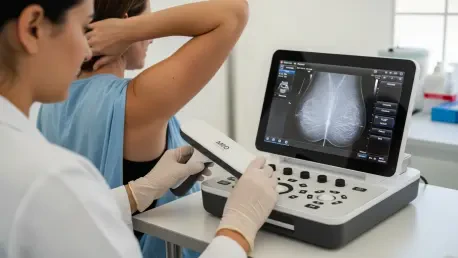

Breast cancer remains a formidable challenge because many aggressive tumors, known as interval cancers, develop rapidly between annual mammograms and often evade detection until they reach advanced stages. Traditional diagnostic workflows rely heavily on centralized hospital infrastructure, which limits the frequency of check-ups for many women and creates a dangerous window of vulnerability for those with fast-growing malignancies. To bridge this gap, engineers at the Massachusetts Institute of Technology have unveiled 3D PURE, a wearable and portable ultrasound system designed to provide laboratory-grade imaging in a decentralized setting. This technology represents a paradigm shift in preventative oncology by allowing for high-resolution, real-time monitoring of breast tissue without the logistical burdens of standard imaging appointments. By utilizing a sophisticated sensor array that can be used more frequently than mammography, this system aims to catch anomalies the moment they appear.
Hardware Evolution: Engineering a Sophisticated Diagnostic Array
At the heart of this portable device is a 128-element two-dimensional transducer array that utilizes a unique “corner-gap offset” geometry to maintain signal integrity in a compact form factor. Standard ultrasound probes often suffer from significant electrical crosstalk, where signals from peripheral elements interfere with one another, leading to saturated data and blurred images. By strategically staggering these elements, the engineering team successfully minimized this interference, allowing the hardware to support higher transmit voltages for deeper tissue penetration. This increased sensitivity is crucial for identifying small masses that might otherwise be obscured by the dense physiological environment of the breast. Consequently, the system can capture high-fidelity volumetric data that rivals much larger stationary units. This mechanical innovation ensures that the device remains both lightweight enough for personal use and powerful enough to satisfy rigorous clinical standards.
Supporting the sensor array is a custom-engineered backing layer that serves a dual purpose as both a mechanical acoustic damper and an electromagnetic shield for the sensitive electronics. Composed of a specialized polyurethane matrix impregnated with tungsten and zirconia particles, this layer is specifically designed to absorb back-propagating ultrasound waves that would otherwise cause ghosting artifacts. By neutralizing these unwanted reverberations, the backing layer significantly increases the device’s operational bandwidth and improves the directionality of the ultrasonic pulses. This results in an exceptionally high signal-to-noise ratio, which is vital for distinguishing between the subtle textures of glandular tissue and the distinct signatures of malignant growths. Furthermore, the shielding properties protect the system from external electromagnetic noise, ensuring that the final image remains crisp even in non-clinical environments. Such material science breakthroughs are what enable the device to maintain its precision across a variety of unpredictable real-world scenarios.
Computational Clarity: Overcoming Tissue Complexity and User Barriers
One of the primary obstacles to accurate breast imaging is the inherent complexity of the anatomy, which contains a heterogeneous mix of fat, fibrous tissue, and glands that causes ultrasound waves to travel at inconsistent speeds. These velocity fluctuations typically lead to phase aberrations, resulting in distorted or low-resolution images that are difficult for radiologists to interpret accurately. To counteract this, the 3D PURE system employs a sophisticated Layered Aberration-Correction Reconstruction algorithm, which performs real-time adjustments to the acoustic beamforming process. By predicting the specific speed-of-sound variations within individual tissue layers, the software can realign the incoming data to produce a coherent and sharp three-dimensional map. This adaptive approach allows the system to maintain high resolution regardless of the patient’s specific tissue density, which has long been a limiting factor for traditional handheld ultrasound devices used in various clinical settings.
Beyond technical imaging challenges, the effectiveness of any handheld diagnostic tool is often limited by the skill of the person operating the probe, a phenomenon known as operator dependence. To democratize the use of high-quality ultrasound, researchers developed the Mirror my First UltraSound interface, which provides real-time visual guidance to ensure the probe is positioned correctly over the target area. This intuitive feedback system allows individuals with no formal medical training to replicate the exact scan parameters used during their initial professional consultation. By matching current scans with historical data overlays, the interface facilitates longitudinal monitoring, making it possible for patients to track specific anomalies from the comfort of home. This breakthrough reduces the need for frequent visits to specialized imaging centers while maintaining a high level of diagnostic consistency over time. Such user-centric design effectively removes the gatekeeping barriers that once restricted advanced imaging to hospitals.
Clinical Validation: Proven Performance and Global Potential
Rigorous performance trials have demonstrated that the 3D PURE system offers a measurable advantage over existing two-dimensional handheld ultrasound alternatives currently available on the market. During comparative testing using anatomically accurate breast phantoms, researchers found that users were able to identify nearly eighty percent of hidden targets when using the 3D system, whereas only sixty percent were caught with standard tools. These findings were further validated through human subject testing, where the device successfully mapped diverse internal structures including cysts, calcifications, and various types of medical implants. The wide volumetric field of view provided by the transducer array ensures that no region is missed during a sweep, providing a comprehensive data set that can be reviewed remotely by oncology experts. This level of reliability is essential for gaining the trust of the medical community and ensuring that portable devices can serve as a dependable first line of defense in cancer detection.
The expansion of this technology into decentralized healthcare models held the potential to significantly improve survival rates by providing underserved populations with access to high-resolution screening. Integrating the hardware with mobile platforms like smartphones and tablets transformed the device into a versatile tool for rural clinics and resource-limited regions where mammography machines were unavailable. Medical professionals recommended that future implementation focused on establishing secure cloud-based pipelines for automated data analysis and specialist consultation to maximize the system’s impact. It became clear that the successful deployment of these portable arrays required a coordinated effort between hardware developers and regional healthcare providers to ensure proper training and follow-up care. By prioritizing accessibility and consistent monitoring, the healthcare industry moved closer to a proactive model of breast health where early detection was no longer a matter of proximity to a major hospital. This shift suggested a new era of personalized diagnostics that empowered patients through technology.