
Diffusion models operate through a sophisticated two-stage process that learns to remove noise and recover original images based on the statistical patterns of high-quality X-ray projections. This development marks a pivotal shift in cone-beam computed tomography (CBCT), a modality that has become indispensable across dental, orthopedic, and oncological centers due to its high spatial resolution. Despite these advantages, the physical dimensions of flat-panel detectors often fail to encompass the entire anatomical volume of larger patients, leading to incomplete data known as truncation. This technical hurdle generates severe artifacts, including bright, bowl-shaped intensity gradients and streaks that obscure critical details. By implementing conditional generative diffusion models (CGDMs), researchers have successfully demonstrated a way to digitally complete this missing information. This approach effectively bypasses the inherent hardware limitations of current scanners, transforming how clinicians interpret 3D data.
Mechanics of Image Distortion
The Physics of Truncation: Mathematical Failures
The foundational mechanism for creating images in CBCT is the Filtered Backprojection algorithm, which relies on the mathematical assumption that every X-ray path passing through an object is captured by the detector. However, when the patient’s anatomy extends beyond the sensor’s field of view, the resulting projection data becomes inherently incomplete. This lack of peripheral information forces the reconstruction algorithm to make erroneous density calculations at the edges of the image. Consequently, the reconstructed volume suffers from the “cupping” effect, where the outer regions appear artificially bright while the central contrast is degraded. In a surgical or diagnostic context, such distortion is more than a mere visual nuisance; it can mask tiny bone fractures or early-stage tumors that reside near the periphery of the scan. This mechanical reality has long necessitated either larger, more expensive hardware or compromised image quality in standard clinical environments.
Beyond the simple loss of spatial data, truncation introduces non-linear errors that propagate throughout the entire reconstructed volume, leading to persistent streaking and intensity non-uniformity. When the X-ray beam interacts with the edges of a patient that the detector cannot see, the missing attenuation values create sharp discontinuities in the mathematical derivative used during the filtering stage of the reconstruction process. These discontinuities manifest as high-frequency noise and shadowing that can alter the Hounsfield units, which are critical for characterizing tissue types. For example, a radiologist attempting to differentiate between various soft tissues in the pelvis or shoulder may find the task impossible if the background intensity is fluctuating due to these mathematical artifacts. As the complexity of modern interventional procedures increases, the need for a more robust mathematical solution that accounts for missing boundary conditions has never been more urgent for patient safety.
Standard Mitigation: Padding and Heuristics
To combat these distortions, medical physicists have historically turned to heuristic methods designed to fill in the gaps in the detector data. The most common technique, integrated into standard platforms like the Reconstruction Toolkit, involves various forms of data padding. This process typically uses simple mirroring of the existing detector pixels or applies smooth mathematical decay functions to bridge the transition between measured data and empty space. While these methods succeed in preventing the most catastrophic reconstruction failures, they do not introduce any new anatomical information. Instead, they provide a smooth boundary that reduces the intensity of the cupping effect without restoring the true structures of the patient. Consequently, the edges of the resulting images remain anatomically inaccurate, providing a cleaner aesthetic but failing to offer the high-fidelity detail required for precision medicine or complex radiation therapy planning.
These traditional approaches also struggle with the inherent variability of human anatomy, as fixed mathematical functions cannot account for the diverse shapes and densities of different patients. Whether the scan involves a muscular shoulder or a wide pelvic girdle, a generic decay function will inevitably fail to match the actual X-ray attenuation that would have occurred. This leads to a persistent mismatch between the padded data and the real anatomy, ensuring that artifacts are merely softened rather than eliminated. Moreover, these manual or semi-automated adjustments often require significant computational overhead or specific calibration for different detector types, complicating the clinical workflow. The limitations of these physics-based shortcuts have highlighted the necessity for a more intelligent, data-driven approach that can infer the likely structure of missing tissues based on prior knowledge of human physiology, leading directly to the current exploration of generative models.
Evolution of Artificial Intelligence Solutions
Adversarial Networks: The Risk of Hallucination
The initial wave of artificial intelligence in medical imaging largely focused on the use of Generative Adversarial Networks for image-to-image translation. These models utilize two competing networks—a generator and a discriminator—to create realistic-looking images from incomplete or noisy data. While these networks were celebrated for their ability to sharpen blurry scans and remove superficial artifacts, they introduced a significant risk into the diagnostic environment: the tendency to hallucinate. Because these models are trained to fool a discriminator into believing an image is real, they may invent anatomical features, such as small vessels or tissue structures, that do not actually exist in the patient. In a clinical setting where every millimeter of data can dictate a surgical path or a cancer diagnosis, such fabrications are unacceptable. This lack of reliability has been a major barrier to the widespread adoption of adversarial solutions in high-stakes medical imaging applications.
Furthermore, the training process for adversarial networks is notoriously unstable, often requiring precise hyperparameter tuning and extensive datasets to avoid issues like mode collapse or inconsistent output quality. This instability means that a model might perform exceptionally well on one patient scan but fail unpredictably on another, depending on how closely the patient’s anatomy matches the training set. For CBCT truncation, where the missing data can vary wildly in shape and volume, the rigidity and unpredictability of adversarial networks pose a significant challenge. Clinicians require a system that is not only visually impressive but also statistically consistent and transparent in how it handles missing data. As researchers sought to move past these limitations, the focus shifted toward diffusion-based architectures, which offer a more controlled and mathematically sound framework for data reconstruction, ensuring that the generated peripheral information remains anchored to the physical reality.
Conditional Diffusion Models: A New Paradigm
Conditional generative diffusion models represent a more sophisticated approach by treating image restoration as a gradual process of noise removal rather than a single-step translation. During the training phase, the model observes how high-quality, full-field X-ray projections are progressively degraded by Gaussian noise until all structural information is lost. It then learns the inverse task, meticulously reconstructing the image by iteratively subtracting noise. When applied to the problem of truncation, the model is conditioned on the existing, truncated projection data. This means the AI does not create an image from scratch; instead, it uses the real measurements as a permanent anchor and intelligently populates the empty peripheral regions with anatomically plausible structures. By maintaining this strict connection to the original sensor data, the diffusion model significantly reduces the probability of hallucinations, ensuring that the generated anatomy matches the statistics.
This methodology effectively merges the predictive power of deep learning with the rigorous requirements of medical physics. By intervening in the projection domain—fixing the raw X-ray data before the 3D volume is reconstructed—the diffusion model allows the standard reconstruction algorithm to function as if it had access to a full-field detector. This shift from post-processing to pre-processing ensures that the final 3D image is free from the underlying mathematical errors that cause cupping and streaking. The iterative nature of the diffusion process also allows for a higher degree of precision, as the model can refine the image at multiple scales, capturing both the broad contours of the patient’s body and the subtle textures of soft tissue. As we move through 2026, this technology is being recognized for its ability to provide a more stable and reliable alternative to previous AI iterations, offering a bridge between traditional imaging science and the next generation of diagnostic tools.
Impact on Clinical Practice
Radiotherapy and Oncology: Target Accuracy
In the specialized field of oncology, the reduction of truncation artifacts is a critical factor for the success of daily radiotherapy sessions. Cancer patients often undergo daily CBCT scans to ensure their body is perfectly aligned with the high-energy radiation beams intended to destroy their tumors. However, truncation artifacts can distort the visible boundaries between a tumor and the surrounding healthy organs, such as the bladder or rectum in prostate cancer treatment. By using conditional diffusion models to clean these images, radiation therapists were able to visualize these interfaces with much greater clarity. This improved visibility directly reduced the risk of positioning errors, ensuring that the radiation dose was concentrated precisely on the intended target while sparing healthy tissue from unnecessary exposure. The enhanced image quality also allowed for more accurate adaptive radiotherapy, where the treatment plan was adjusted in real-time.
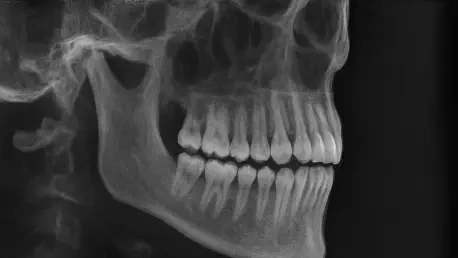
Beyond radiotherapy, the enhanced diagnostic reliability provided by diffusion models has transformed dental and oral surgery practices. Dental CBCT units often utilize very small fields of view to minimize the radiation dose to the patient’s head and neck. However, this focus frequently results in the truncation of the jawbone, sinuses, or airway, which are vital for planning dental implants or corrective jaw surgeries. The integration of AI-driven reconstruction enabled surgeons to view the entire maxillofacial structure with uniform clarity, eliminating the distortions that typically occur at the detector’s edge. This allowed for more precise surgical planning and reduced the need for repeated scans, which further limited the patient’s radiation exposure. By providing a complete anatomical context within a limited scan, the diffusion model effectively maximized the clinical utility of every X-ray projection, making complex oral procedures safer and more predictable.
Future Implementation: Scalability and Access
The successful demonstration of conditional generative diffusion models in reducing CBCT truncation artifacts established a new benchmark for software-based image enhancement. Researchers concluded that the transition from physics-based heuristics to data-driven AI significantly improved the accuracy of 3D reconstructions. To build on this foundation, future efforts were directed toward the integration of these algorithms into real-time imaging workflows, ensuring that the noise-removal process occurred within the seconds between the scan and the clinician’s review. Large-scale clinical trials were initiated to validate the model’s performance across a wider variety of detector technologies and patient populations to ensure universal applicability. Healthcare providers were encouraged to adopt these AI protocols as a standard enhancement to their existing CT infrastructure, ensuring that hardware limits no longer dictated the quality of care.
By prioritizing data fidelity over simple visual smoothing, the medical community took a decisive step toward a future where hardware limits no longer dictated the quality of patient care. The researchers recommended that manufacturers begin embedding these diffusion models directly into the firmware of new CBCT units to provide seamless, artifact-free imaging out of the box. Furthermore, the development of lighter, more computationally efficient versions of these models was prioritized to allow for deployment on mobile and point-of-care imaging devices. As these systems became more widespread, the focus shifted toward longitudinal studies to determine the long-term impact on patient survival rates and surgical success. This progression ensured that the technological breakthrough translated into tangible improvements in global health outcomes, solidifying the role of generative AI as a permanent fixture in the modern radiology suite for years to come.