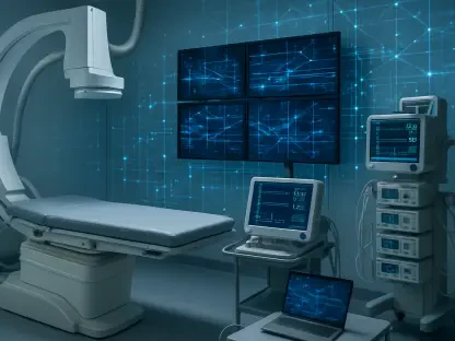

Scientists at the National Ignition Facility have successfully transformed the world’s most energetic laser into a diagnostic tool capable of producing the most brilliant X-ray pulses ever recorded in a laboratory setting. While the facility is primarily known for achieving fusion ignition, the recent refinement of the Advanced Radiographic Capability has introduced a new paradigm in high-energy-density physics. This sophisticated system allows researchers to bypass the limitations of traditional imaging by generating mega-electron-volt X-rays that can pierce through materials previously thought to be opaque. By concentrating massive amounts of energy into snapshots lasting only a few trillionths of a second, the system provides a visual gateway into the heart of extreme physical reactions. This capability is not merely a technical curiosity but a fundamental necessity for understanding how matter behaves when compressed to densities found only in the cores of stars or within the fleeting moments of a nuclear detonation. This progress represents the culmination of a multi-year effort to refine diagnostic precision and energy delivery within the chamber.
The Engineering: Mechanics of the Advanced Radiographic Capability
The operational brilliance of the Advanced Radiographic Capability lies in its ability to modify the existing infrastructure of the primary laser system to perform a specialized secondary function. By strategically compressing two of the facility’s primary beamlines, the system delivers kilojoules of energy within a span of mere picoseconds, making it the most energetic short-pulse laser currently in operation. This rapid delivery of power is essential for creating high-energy X-rays that possess enough penetrating force to visualize dense materials under pressure. Unlike standard radiographic methods that may struggle with the extreme background noise of a fusion experiment, this laser-driven approach generates a signal so intense that it stands out clearly against the surrounding radiation. The process effectively turns the laser into a high-speed camera, capable of freezing the motion of particles moving at hyper-velocity. This technical feat required years of modeling and hardware adjustments to ensure the beamlines could be synchronized with the precision necessary for scientific validity.
This engineering achievement facilitates the creation of mega-electron-volt X-rays, which are significantly more powerful than those used in medical or industrial imaging. The primary objective is to observe the hydrodynamic behavior of materials as they are subjected to intense shockwaves. When the short-pulse laser strikes a specific target, it triggers a cascade of high-energy electrons that subsequently produce a burst of X-rays. This burst is what allows physicists to see through several centimeters of dense metals like lead or tungsten, which would normally block any form of light or lower-energy radiation. The development of this capability has effectively expanded the diagnostic toolkit available for high-energy experiments, providing a way to verify theoretical models of material strength and fluid dynamics. As researchers continue to push the boundaries of laser intensity, the ability to maintain the stability and focus of these pulses remains a top priority for the engineering teams involved. This ensures that every experiment yields high-fidelity data that can be used to inform broader scientific conclusions.
The Detection Challenge: Quantifying Brightness and Precision
One of the most significant hurdles in developing high-energy radiography is the paradoxical nature of the X-rays themselves, as their extreme penetrating power makes them difficult to capture. Because these photons pass through most substances with ease, they also tend to pass through standard diagnostic sensors without leaving a measurable trace or depositing enough energy for a clear reading. To solve this problem, a specialized suite of advanced diagnostics was integrated into the experimental setup, combining gamma reaction history tools with electron-proton-positron spectrometers. These instruments allowed the research team to finally quantify the “brightness” and “source size” of the generated X-rays. In the realm of experimental physics, brightness refers to the total number of photons produced in a single burst, while the source size determines the ultimate sharpness of the resulting image. Achieving a balance between a high photon count and a microscopic source area is what allows for the production of high-resolution radiographs that can resolve internal features.
The refinement of these measurement techniques has allowed for a much deeper understanding of how the laser energy is converted into radiographic light. By deploying nuclear activation diagnostics alongside traditional sensors, the team was able to map the energy distribution of the X-ray pulses with unprecedented accuracy. This data was crucial for optimizing the target designs and laser configurations used in subsequent experiments. The research indicated that the highest quality images were consistently obtained when the objects were positioned precisely along the laser axis line-of-sight. This discovery led to the development of specialized sample holders and diagnostic frames designed to maintain sub-millimeter alignment within the massive target chamber. This level of precision is necessary because even a minor deviation in the positioning of the target or the detector could result in a blurred image, rendering the data useless for detailed analysis. The successful integration of these diagnostics has turned a difficult measurement task into a repeatable and reliable scientific process.
Experimental Validation: Piercing through Dense Materials
To demonstrate the practical utility of this ultra-bright X-ray source, researchers conducted a series of rigorous tests using targets specifically designed to challenge the limits of modern imaging. These experiments utilized small lead spheres featuring internal ripples and structures measured in the hundreds of microns, which were then placed behind thick layers of tungsten. Both lead and tungsten are exceptionally dense, representing the most difficult materials for any radiographic system to penetrate. The results were definitive, as the system successfully captured clear images of the internal ripples even through the heavy shielding of the tungsten filters. This proved that the laser-based X-ray source could resolve fine internal structures in environments that would be completely inaccessible to conventional flash X-ray technology. The clarity of these radiographs provided visual confirmation that the system could be used to monitor the evolution of complex shapes and material interfaces during the high-speed motion characteristic of hydrodynamic events.
The successful validation of these high-energy sources established a clear path toward even more ambitious goals in the coming years. Following the completion of the initial testing phase, the research team finalized a roadmap to improve spatial resolution from its current levels down to approximately 10 microns. This transition involved a collaborative effort between multiple national laboratories and academic institutions, ensuring that the technology remained at the forefront of the scientific community. The project participants documented the scaling of these X-ray sources and integrated the findings into the broader framework of national security and fundamental nuclear research. By demonstrating that laser-driven radiography could provide better detail than existing methods, the initiative secured its role as a critical component of future high-energy-density experiments. The move toward higher resolution promised to unlock new insights into the microscopic behavior of matter under stress, providing a more granular look at the physics of the extreme. All milestones reached during this period served to solidify the facility’s reputation as a premier site for optical and nuclear science.