
Advanced medical imaging is now offering an unprecedented, non-destructive window into the distant past, allowing researchers to diagnose illnesses in individuals who lived and died more than two millennia ago. Through a remarkable fusion of radiology and archaeology, scientists are digitally unwrapping ancient Egyptian mummies, revealing that the physical complaints of the ancient world are strikingly similar to those faced by people today. A recent project focusing on two priests, Nes-Min, who lived around 330 BCE, and Nes-Hor, from approximately 190 BCE, has utilized high-resolution computed tomography (CT) to peer beneath their linen wrappings. This technological approach not only preserves the sanctity and physical integrity of these fragile human remains but also generates a wealth of data that reconstructs their health profiles with astonishing clarity. The findings underscore a profound connection across time, demonstrating that fundamental human health challenges, from degenerative joint disease to dental problems, are a timeless aspect of the human condition.
A Digital Glimpse into Ancient Lives
The Power of Non-Invasive Exploration
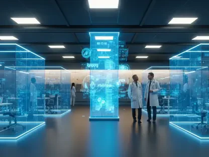
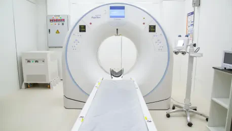
The core innovation driving this historical investigation is the application of high-resolution CT scanning, a technology that enables a form of “digital unwrapping.” This sophisticated process allows scientists to meticulously examine the internal structures of the mummies without physically disturbing their delicate wrappings or the remains within. The scanners capture thousands of cross-sectional images, which are then compiled by powerful computers to create comprehensive, three-dimensional models. This method yields an extraordinary level of detail, making it possible to inspect the skeletal system for fractures and signs of disease, identify remnants of soft tissue, and even discern subtle facial features like eyelids and lips, all while the individuals remain securely sealed within their sarcophagi. This non-destructive technique represents a monumental leap forward from earlier archaeological methods, which often required damaging or destroying the very artifacts being studied. It ensures the preservation of these invaluable historical subjects for future generations of researchers while providing a more complete and intimate understanding of their lives.
This advanced imaging workflow directly mirrors cutting-edge practices in contemporary clinical medicine, where surgeons frequently rely on patient-specific 3D models to plan intricate and high-risk procedures. By applying this same methodology to archaeological subjects, researchers can not only diagnose ancient pathologies but also create tangible, physical replicas of bones and artifacts using 3D printing technology. This synergy between medical science and historical inquiry is transformative. The ability to produce life-size, anatomically precise models allows for hands-on study and analysis that would otherwise be impossible. It bridges the gap between digital data and physical reality, enabling a deeper appreciation of the anatomical challenges these ancient individuals faced. This dual application highlights the versatility of modern imaging, demonstrating how a single technological platform can simultaneously advance both patient care in the present and our collective understanding of human history, providing insights that were once confined to the realm of speculation.
Timeless Pathologies Revealed
The detailed scans of the two Egyptian priests, Nes-Min and Nes-Hor, have unearthed compelling evidence of ailments that resonate strongly with modern medical diagnoses. Nes-Min, who is believed to have died at a younger age, was found to have a collapsed lumbar vertebra, a condition indicative of age-related degeneration that is commonly seen in patients today. This discovery challenges the notion that such wear-and-tear conditions are solely a product of modern lifestyles or longer lifespans. Meanwhile, Nes-Hor, who lived to an older age, exhibited signs of more advanced physical decline. His scans revealed significant deterioration in his hip joint, a pathology consistent with severe arthritis that would have likely caused him chronic pain and limited his mobility. Furthermore, he suffered from extensive dental problems, including abscesses and missing teeth, another common complaint in both ancient and modern populations. These specific, relatable health issues paint a vivid picture of the physical burdens carried by individuals in the ancient world, humanizing them in a way that historical texts alone cannot.
The findings from these digital autopsies contribute to a growing consensus among historians and medical experts that many fundamental human health challenges are universal and have persisted across millennia. The presence of degenerative bone disease, joint deterioration, and poor dental health in these ancient priests demonstrates that these were not isolated issues but common afflictions that transcended time and culture. By providing clear, visual evidence of these conditions, the CT scans offer a powerful counter-narrative to romanticized views of the past. Life in ancient Egypt, even for esteemed individuals like priests, involved tangible physical suffering that is all too familiar to the modern world. This shared biological heritage connects us directly to these distant ancestors, showing that despite vast differences in technology, culture, and environment, the human body has always been susceptible to the same fundamental vulnerabilities and processes of aging and decay.
A New Era for Historical Inquiry
The Convergence of Disciplines
The successful analysis of Nes-Min and Nes-Hor exemplifies a powerful and growing trend: the convergence of medical technology and historical research. This interdisciplinary approach is reshaping the field of archaeology, moving it beyond traditional excavation and analysis toward a more holistic and data-driven science. Radiologists, with their expertise in interpreting complex medical images, are now collaborating closely with Egyptologists and historians to unlock secrets that have been sealed away for centuries. This synergy allows for a much deeper and more nuanced interpretation of ancient remains. While an archaeologist can identify the context and cultural significance of a mummy, a radiologist can diagnose specific diseases, estimate age at death with greater accuracy, and even infer aspects of an individual’s lifestyle and diet based on skeletal evidence. This collaborative model harnesses the strengths of multiple fields to construct a more complete and scientifically rigorous picture of the past, transforming ancient mummies from silent artifacts into detailed case studies of human health and history.
The public exhibition of these findings marks a crucial step in making this specialized scientific discovery accessible and engaging for a broader audience. By displaying the mummies alongside their detailed digital reconstructions and 3D-printed skeletal models, institutions like the California Science Center are translating complex scientific data into a compelling and understandable narrative. This approach allows visitors to see for themselves the evidence of ancient diseases and to appreciate the sophisticated technology used to uncover them. It demystifies the research process and fosters a greater public appreciation for the intersection of science, history, and medicine. Making this knowledge accessible not only educates the public but also inspires a new generation of scientists and historians, demonstrating the exciting possibilities that emerge when different fields of expertise unite to explore the enduring questions of human existence and our shared past.
Looking to the Past to Understand the Present
This investigation ultimately provided a profound perspective on the continuity of the human experience. The research on the mummies of Nes-Min and Nes-Hor did more than just identify ancient ailments; it reinforced the understanding that the biological challenges of life have remained remarkably consistent through the ages. The project successfully demonstrated how modern clinical workflows could be adapted to serve historical inquiry, establishing a powerful precedent for future archaeological studies. By bridging the gap between ancient history and modern medicine, these digital explorations offered a humanizing glimpse into the lives of individuals who lived millennia ago, reminding us of our shared vulnerability to pain, disease, and the inevitable process of aging. The knowledge gained from their preserved remains created a tangible link to the past that continues to inform our understanding of human health today.