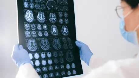
In the realm of medical diagnostics, brain tumor identification remains a formidable challenge, with millions of patients worldwide relying on precise imaging for timely treatment, and Magnetic Resonance Imaging (MRI) serves as a cornerstone in this process. However, the complexity of tumor shapes, sizes, and locations often complicates accurate segmentation and classification. Enter PolicySegNet, a groundbreaking hybrid deep learning framework that promises to transform this landscape by integrating advanced transformer-based feature extraction with reinforcement learning. Designed to simultaneously segment and classify brain tumors in MRI scans, this model leverages a pretrained SegFormer-B4 encoder, a UNet-inspired decoder, and a lightweight classification head. Its use of proximal policy optimization (PPO) to balance both tasks through a reward system marks a significant departure from traditional methods. With impressive performance metrics—segmentation accuracy nearing 99% and classification accuracy up to 91.75% on validation sets—PolicySegNet offers a glimpse into a future where AI could enhance clinical decision-making, potentially reducing diagnostic errors and improving patient outcomes. This article explores the intricacies of this innovative tool, its methodology, and its potential to reshape brain tumor diagnosis.
1. Understanding the Challenges in Brain Tumor Diagnosis
Brain tumors present a critical challenge in medical imaging due to their diverse morphologies and often indistinct boundaries, which can obscure precise identification in MRI scans. Accurate segmentation—delineating the exact tumor area—and classification—determining the tumor type—are essential for effective treatment planning, yet these tasks are fraught with difficulties. Variability in tumor appearance, coupled with surrounding edema or tissue distortion, frequently complicates the process for radiologists. Traditional deep learning approaches, predominantly based on convolutional neural networks (CNNs), have shown promise but often fall short in capturing long-range dependencies within complex medical images. This limitation can result in missed or inaccurate diagnoses, particularly for subtle or overlapping tumor features, underscoring the need for more advanced methodologies that can adapt to such intricacies.
The computational burden of fine-tuning large-scale models for specific medical tasks adds another layer of difficulty, especially when labeled data is scarce. Many existing frameworks require extensive datasets to achieve reliable performance, a luxury not always available in clinical settings. Moreover, balancing segmentation accuracy with classification precision in a unified system remains an elusive goal for most models. PolicySegNet emerges as a potential solution to these persistent issues, aiming to address both tasks efficiently. By combining cutting-edge transformer technology with a novel reinforcement learning strategy, it seeks to overcome the shortcomings of conventional tools, paving the way for more reliable and resource-efficient diagnostics in neuro-oncology.
2. Introducing PolicySegNet’s Innovative Framework
PolicySegNet represents a paradigm shift in brain tumor analysis by integrating a hybrid deep learning architecture tailored for joint segmentation and classification of MRI scans. At its core lies a pretrained SegFormer-B4 encoder with a Mix Transformer (MiT) backbone, originally trained on a large dataset for general image segmentation. This encoder remains fixed during training, serving as a robust feature extractor that captures both local and global image details without the need for computationally expensive fine-tuning. Paired with a UNet-inspired decoder for segmentation and a lightweight classification head for tumor type identification, the framework achieves efficiency while tackling the dual objectives of precise localization and accurate diagnosis.
A distinguishing feature of this model is its use of proximal policy optimization (PPO), a policy-based reinforcement learning algorithm. PPO optimizes the decoder and classifier simultaneously through a balanced reward signal that prioritizes both segmentation accuracy and classification performance. This approach contrasts with traditional supervised learning methods by dynamically adjusting to task-specific challenges, such as class imbalance or ambiguous tumor boundaries. With reported segmentation accuracies as high as 0.9961 on training data and classification accuracies reaching 0.9175 on validation sets, the framework demonstrates remarkable potential. Its ability to operate effectively on limited domain-specific data further enhances its applicability in real-world clinical environments.
3. Delving into the Transformer Encoder and Feature Extraction
The transformer encoder in PolicySegNet, based on the SegFormer-B4 architecture, employs a hierarchical Mix Transformer (MiT) design to extract features from MRI scans with exceptional detail. This process begins with overlapping patch embedding, where input images are divided into patches using a convolutional layer with specific stride, kernel size, and padding parameters. This ensures preservation of fine-grained details while reducing spatial dimensions into feature maps. Multi-Head Self-Attention (MSA) mechanisms then process these embeddings to model global dependencies, allowing the encoder to capture relationships across the entire image rather than just localized areas, a significant advantage over traditional CNNs in medical imaging tasks.
Following attention processing, a mix-feed forward network (Mix-FFN) refines the features by integrating linear projections with depth-wise convolutions for local context modeling. The hierarchical structure operates through four stages, progressively reducing spatial resolution while increasing feature dimensionality to capture both fine and coarse details. Residual connections and layer normalization at each step ensure stable training and robust feature representation. The resulting multi-scale embeddings are then passed to the UNet-based decoder for segmentation. This sophisticated encoder design enables PolicySegNet to efficiently discern intricate tumor boundaries and contextual information, setting a strong foundation for accurate diagnostic outputs in complex MRI analyses.
4. Exploring the UNet Decoder for Segmentation
The UNet decoder within PolicySegNet plays a pivotal role in reconstructing detailed segmentation masks from the hierarchical embeddings provided by the transformer encoder. This component employs a progressive up-sampling strategy using transposed convolutions to increase the spatial resolution of feature maps. At each stage, a transposed convolution operation with defined stride, kernel size, and padding enhances the dimensions, followed by batch normalization and ReLU activation to stabilize and refine the output. This initial step ensures that the model can scale up low-resolution features while maintaining critical spatial information necessary for precise tumor delineation.
Subsequent refinement occurs through additional convolutional layers with ReLU activation to restore fine details and enhance non-linearity, crucial for capturing subtle tumor edges. The decoder comprises five transposed convolution blocks, each progressively increasing resolution to align with the original input dimensions. The final segmentation mask is generated using a 1×1 convolution to map features to the required output channels, followed by a sigmoid activation for binary segmentation probabilities. This lightweight yet effective architecture requires fewer parameters compared to deeper decoders, ensuring computational efficiency. By prioritizing detailed segmentation through hierarchical up-sampling and edge refinement, the UNet decoder significantly contributes to PolicySegNet’s high segmentation accuracy across varied tumor types.
5. Unpacking the Classification Head for Tumor Identification
The classification head in PolicySegNet is engineered to predict tumor types from the extracted feature embeddings, complementing the segmentation task. Initially, global average pooling (GAP) is applied to reduce each feature map’s spatial dimensions by averaging values across height and width, resulting in a compact vector representation per channel. This process effectively summarizes spatial information, serving as a lightweight alternative to traditional fully connected layers, and prepares the data for efficient classification without excessive computational overhead. The streamlined approach ensures that critical diagnostic features are retained for accurate tumor categorization.
Following this reduction, the flattened feature vector is processed through a fully connected layer that maps it to the number of tumor classes, specifically four in this case: No Tumor, Glioma, Meningioma, and Pituitary. During inference, a softmax activation function converts the output logits into class probabilities, enabling clear identification of the most likely tumor type. This design prioritizes efficiency, allowing the classification head to operate with minimal resource demands while achieving robust performance. By integrating seamlessly with the segmentation pipeline, this component ensures that PolicySegNet delivers comprehensive diagnostic insights, aligning spatial mapping with categorical predictions for enhanced clinical utility.
6. Leveraging PPO for Reinforcement Learning Optimization
PolicySegNet’s integration of proximal policy optimization (PPO) marks a significant advancement in optimizing joint segmentation and classification tasks through reinforcement learning. Unlike traditional policy gradient methods, PPO employs a clipped surrogate objective to prevent large policy updates, ensuring stability during training. It alternates between collecting trajectories with the current policy and updating it based on advantage estimates, balancing exploration with reliable learning. This methodology is particularly effective for complex tasks like medical image analysis, where balancing multiple objectives is crucial for accurate outcomes.
In this framework, the PPOAgent orchestrates optimization by combining supervised losses with a reinforcement learning reward signal. The forward pass involves feature extraction via the SegFormer encoder, segmentation prediction through the UNet decoder, and tumor type classification using averaged embeddings. The reward function evaluates segmentation quality via intersection over union (IoU) and classification accuracy, merging these metrics into a unified score. Total loss integrates supervised segmentation and classification losses with the scaled RL reward, guiding model updates through backpropagation using the Adam optimizer. This dual-learning approach mitigates issues like class imbalance and boundary ambiguity, enhancing PolicySegNet’s precision and reliability in clinical diagnostics.
7. Examining Experimental Settings and Dataset Composition
The experimental foundation of PolicySegNet rests on a comprehensive dataset of 4238 MRI brain scans, sourced publicly and resized to 512×512 pixels for consistency. These images are categorized into four classes—No Tumor, Glioma, Meningioma, and Pituitary—each accompanied by binary segmentation masks for precise pixel-wise annotation. To ensure robust evaluation, the dataset is split into training (80%, 3390 images), validation (10%, 424 images), and testing (10%, 424 images) subsets. This stratified division facilitates effective learning, hyperparameter tuning, and unbiased assessment of the model’s performance across diverse tumor scenarios.
Training for PolicySegNet spans 40 epochs, utilizing the Adam optimizer with a learning rate of 0.0001 to refine the trainable parameters of the UNet decoder and classification head. The fixed SegFormer-B4 encoder minimizes computational demands, allowing focus on task-specific optimization. This setup ensures that the model adapts efficiently to the limited yet critical medical imaging data, a common constraint in clinical environments. By structuring the experiment to mirror real-world diagnostic challenges, including varied tumor types and image distributions, the settings provide a solid basis for evaluating PolicySegNet’s potential impact on brain tumor analysis workflows.
8. Analyzing Performance Results and Comparative Insights
PolicySegNet’s performance metrics reveal exceptional capabilities in brain tumor diagnosis, with segmentation accuracy consistently around 99% across training, validation, and test phases. Classification accuracy also stands out, reaching up to 91.75% on validation data, particularly excelling in identifying non-tumor cases and high-grade tumors like pituitary tumors. Class-specific metrics indicate strong true positive rates for No Tumor and Pituitary classes, though lower sensitivity for Glioma reflects challenges in under-segmentation rather than misclassification. These results highlight the model’s ability to handle biological heterogeneity and indistinct tumor margins often encountered in neuro-oncological imaging.
When compared to a non-PPO variant, PolicySegNet demonstrates superior F1-scores for both segmentation and classification, alongside improved segmentation quality with more coherent tumor boundaries. Against recent studies, such as the work by Akter et al. in 2024, this model showcases advantages in joint task optimization through a unified single-stage approach, despite a slightly lower test classification accuracy of 88.03% compared to 97.7%. The use of PPO enhances generalization and stability, reducing high-risk errors critical in medical contexts. These comparative insights affirm PolicySegNet’s strength in delivering reliable, clinically meaningful results, positioning it as a formidable tool in diagnostic innovation.
9. Reflecting on Broader Impacts and Future Potential
The broader implications of PolicySegNet extend far beyond technical achievements, offering transformative potential for brain tumor diagnosis and treatment planning. By efficiently operating on limited datasets with a frozen encoder, the framework becomes accessible to resource-constrained settings, such as smaller clinics lacking high-performance computing infrastructure. Its high accuracy across multiple tumor types enhances clinical decision-making, potentially enabling earlier interventions and reducing diagnostic errors. This could directly translate into improved patient outcomes, particularly in regions where advanced diagnostic tools are scarce.
Looking ahead, PolicySegNet sets a precedent for developing more generalizable AI tools in medical imaging, fostering collaboration between technology researchers and healthcare professionals. Its interpretable design and reinforcement learning approach could inspire similar innovations across other medical domains, addressing complex diagnostic challenges. As deployment in real-world settings progresses, the model’s ability to support safer surgical planning and personalized treatment strategies might democratize access to cutting-edge medical technology. Ultimately, this framework stands poised to contribute significantly to global health equity, ensuring that advanced diagnostics are not confined to well-resourced environments but reach patients universally.


