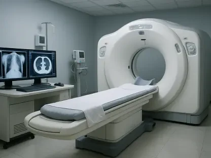

In a world where medical diagnostics are becoming increasingly critical for early detection and personalized treatment, a groundbreaking innovation from the UC Davis Department of Radiology is capturing attention with its potential to revolutionize the field. Supported by a substantial $2.5 million grant from the National Institutes of Health over four years, researchers have developed a hybrid imaging technique known as PET-enabled Dual-Energy CT. This method merges the strengths of Positron Emission Tomography (PET) and dual-energy Computed Tomography (CT) to deliver unprecedented detail in medical scans. By enhancing the ability to detect and characterize diseases like cancer, bone marrow disorders, and heart disease, this technology promises to redefine diagnostic precision without increasing radiation exposure or requiring new equipment. The potential to transform patient outcomes through sharper, more insightful imaging is sparking excitement across the medical community, raising questions about how soon this could become a standard in clinical practice.
Breaking New Ground in Diagnostic Imaging
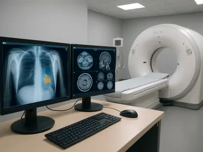
The core innovation of PET-enabled Dual-Energy CT lies in its ability to combine two established imaging modalities into a single, powerful tool. Traditional PET/CT scans, often used in oncology, rely on single-energy CT, which struggles to distinguish between different tissue types. In contrast, this new approach utilizes PET data to generate a high-energy CT image alongside the standard scan, creating a dual-energy perspective. This fusion offers a clearer view of tissue composition, allowing clinicians to not only pinpoint abnormalities but also understand their nature. For instance, distinguishing between healthy and malignant tissues becomes more precise, potentially leading to earlier diagnoses. Such advancements could significantly alter the trajectory of patient care by providing deeper insights into complex conditions without the need for multiple tests or invasive procedures, streamlining the diagnostic process.
Beyond its technical prowess, this hybrid imaging technique addresses a critical need for enhanced accuracy across various medical fields. In cancer detection, the improved differentiation of tissues can mean the difference between timely intervention and delayed treatment. For bone marrow disorders, such as leukemia, the technology provides a more detailed assessment of disease activity, which is vital for monitoring progression and response to therapy. Additionally, in the realm of cardiology, it offers potential insights into the links between bone marrow activity, systemic inflammation, and cardiovascular risks. By integrating metabolic data from PET with structural details from CT in one scan, this method enhances tumor characterization and supports more tailored treatment plans. The breadth of its applications underscores its potential to become a cornerstone of modern diagnostics, pushing the boundaries of what imaging can achieve.
Accessibility and Cost-Effectiveness as Game Changers
One of the most compelling aspects of PET-enabled Dual-Energy CT is its compatibility with existing medical infrastructure. Unlike many cutting-edge technologies that require costly hardware upgrades, this technique can be implemented on numerous current PET/CT scanners with minimal adjustments. This accessibility addresses a significant barrier in adopting advanced imaging solutions, making it feasible for a wider range of hospitals and clinics to offer state-of-the-art diagnostics. Patients stand to benefit from this cost-effectiveness, as the financial burden of accessing such technology is reduced. The research, conducted using the innovative EXPLORER total-body PET scanner, has shown promising results, indicating that the integration of this method into routine practice could be closer than expected, democratizing high-quality care.
Furthermore, the economic implications of this technology extend beyond individual healthcare facilities to the broader medical landscape. By leveraging existing equipment, the adoption of PET-enabled Dual-Energy CT minimizes the need for substantial capital investments, which often delay the rollout of new diagnostic tools. This practicality is especially crucial for smaller or underfunded institutions that struggle to keep pace with technological advancements. The ability to enhance diagnostic capabilities without prohibitive costs could lead to more equitable healthcare delivery, ensuring that patients in diverse settings receive the benefits of precise imaging. As ongoing studies continue to validate its effectiveness, the medical field anticipates a shift toward broader implementation, potentially reshaping how resources are allocated for diagnostic innovations in the coming years.
Collaborative Efforts and Future Validation
The development of this transformative imaging technology is a testament to the power of collaboration within the medical research community. A dedicated team from the UC Davis Department of Radiology and the UC Davis Comprehensive Cancer Center, led by Professor Guobao Wang, has driven this project forward with a shared vision of improving patient outcomes. Their efforts, bolstered by prior funding from a Trailblazer R21 Award from the National Institutes of Health, reflect a commitment to rigorous testing and refinement. The focus now lies on ensuring the reliability of PET-enabled Dual-Energy CT in real-world clinical settings, a critical step before it can be widely adopted. Such collaborative endeavors highlight the importance of interdisciplinary expertise in tackling complex challenges in medical imaging.
Equally important is the ongoing commitment to validating this technology through comprehensive clinical trials. While initial results are promising, as evidenced by recent publications in respected journals, further research is essential to confirm its consistency across diverse patient populations and conditions. The research team is actively working to address potential limitations and optimize the technique for practical use. This phase of development is crucial for building confidence among healthcare providers who will ultimately integrate this tool into their diagnostic arsenal. By prioritizing thorough evaluation, the project aims to establish a robust foundation for future applications, ensuring that the benefits of enhanced imaging translate into tangible improvements in patient care over time.
Paving the Way for Precision Medicine
Reflecting on the journey of PET-enabled Dual-Energy CT, it’s evident that the strides made by the UC Davis team mark a significant milestone in medical imaging history. The fusion of PET and dual-energy CT into a seamless diagnostic tool tackles longstanding challenges in tissue differentiation and disease characterization with remarkable ingenuity. Its ability to provide detailed insights into conditions like cancer, bone marrow disorders, and heart disease sets a new standard for accuracy, while its compatibility with existing scanners ensures widespread potential impact. Looking ahead, the focus shifts to actionable next steps, including expanded clinical testing and integration into standard protocols. The medical community eagerly anticipates partnerships with technology providers to streamline adoption, alongside efforts to train clinicians on its use. These initiatives promise to solidify this innovation as a cornerstone of precision medicine, ultimately enhancing patient outcomes through tailored, data-driven care.