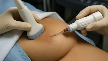

The recommendation for a breast biopsy can trigger a cascade of anxiety and uncertainty, a difficult experience made more common by the limitations of current screening technologies. For many women, particularly those with dense breast tissue, standard mammograms and follow-up ultrasounds often produce ambiguous images, leading to a high rate of false positives where benign cysts are flagged as potentially cancerous. This diagnostic gray area results in millions of invasive procedures that ultimately prove to be unnecessary, creating significant emotional and physical burdens for patients. In an effort to address this long-standing challenge, a team of researchers has developed a groundbreaking ultrasound technology. This innovation, rooted in a novel signal-processing method rather than new hardware, promises to deliver unprecedented clarity, potentially transforming the standard of care by significantly reducing the need for biopsies and alleviating patient distress from the very first scan.
Understanding the Diagnostic Challenge
The Issue with Dense Breast Tissue
The primary obstacle in accurately diagnosing breast abnormalities via conventional ultrasound lies in a phenomenon known as “acoustic clutter,” which is especially prevalent in women with dense breast tissue. In this environment, sound waves do not travel cleanly; instead, they scatter as they move through the tissue, creating significant noise and interference in the resulting image. This scattering effect can profoundly distort the visual characteristics of a breast mass. A benign, fluid-filled cyst, which should ideally appear as a distinct, black void on an ultrasound, becomes obscured by this acoustic clutter. The clutter causes the cyst to appear gray and gives it a visual texture that is nearly indistinguishable from that of a solid, potentially cancerous mass. This profound ambiguity forces radiologists to operate with an abundance of caution. Faced with an image that could represent either a harmless cyst or a malignant tumor, the standard protocol often necessitates recommending a biopsy to achieve a definitive diagnosis, contributing to the high volume of procedures performed on benign lesions.
Introducing Coherence-Based Imaging
The technological breakthrough from Johns Hopkins confronts this problem by fundamentally reimagining how ultrasound data is interpreted. Traditional ultrasound systems create images based on the amplitude, or strength, of the returning sound wave signals. The new approach, however, employs a “coherence-based” method that analyzes the similarity and consistency between a given signal and its immediate neighbors. By prioritizing the uniformity of the returning sound waves, the system’s software can intelligently distinguish between the coherent signals that form a true anatomical structure and the random, incoherent signals that constitute acoustic clutter. This sophisticated filtering process effectively strips away the visual noise, producing a significantly cleaner and higher-contrast image. The result is a clear and unambiguous depiction of the mass, allowing for a reliable and confident differentiation between the smooth, well-defined borders of a fluid-filled cyst and the more complex structure of a solid mass, directly addressing the core issue that drives false positives.
The Impact on Clinical Practice and Patient Care
A Leap in Diagnostic Accuracy
The clinical efficacy of this innovative coherence-based imaging method has been demonstrated through compelling results from an initial study involving 132 patients. The findings revealed a dramatic enhancement in diagnostic precision. When utilizing the new technology, radiologists were able to correctly identify the nature of breast masses—distinguishing between benign cysts and solid masses—with an accuracy rate of 96%. This figure stands in stark contrast to the 67% accuracy rate achieved by the very same group of doctors when they analyzed the exact same set of masses using their standard, amplitude-based ultrasound equipment. This substantial 29-percentage-point improvement represents a monumental leap forward in diagnostic capability. It validates the technology not merely as an incremental upgrade but as a transformative tool that can provide a much higher degree of certainty at a critical stage of the screening process, greatly improving the reliability of the initial assessment and forming a stronger foundation for subsequent clinical decisions.
Beyond a Clearer Picture Aiding Decision-Making
This advanced technology elevates the diagnostic process beyond simply providing a clearer image; it introduces an objective, quantitative component that supports clinical decision-making. The system analyzes each detected mass and generates a numerical score. A specific, predetermined threshold is set, and only masses that score above this threshold are flagged as suspicious and potentially warranting further investigation. This feature helps to automate a complex and often subjective interpretive task, thereby reducing what is known as “decision fatigue” for radiologists who review numerous scans daily. By providing a data-driven metric, the technology allows clinicians to make diagnoses with a much higher level of immediate confidence. The direct benefit for patients is profound. This increased certainty at the point of care can significantly decrease the number of false-positive results, which in turn reduces the need for stressful follow-up appointments, painful biopsies, and the prolonged emotional toll associated with diagnostic uncertainty.
Looking Ahead The Technology’s Future Potential
From AI Integration to At-Home Screening
Looking forward, the research team identified a powerful synergy between this coherence-based imaging and the rapidly advancing field of artificial intelligence. The superior image quality produced by the new system could serve as a much cleaner and more reliable input for AI platforms that are already being trained to distinguish between benign and malignant masses. By feeding these advanced algorithms higher-quality data, the combined system could create a formidable diagnostic tool capable of rendering a highly accurate assessment of a mass’s composition and cancer risk in a single, initial appointment. This integration held the potential to streamline the entire diagnostic workflow, providing clinicians and patients with definitive answers faster than ever before. The fusion of enhanced imaging with intelligent analysis promised a future where the path from detection to diagnosis would be significantly shortened and far more precise.
Democratizing Breast Health Monitoring
The long-term vision for this technology extended beyond the clinical setting, aiming to democratize breast health monitoring for the general population. As ultrasound technology was projected to become more affordable and accessible, the researchers envisioned the integration of the coherence-based method into inexpensive, user-friendly, at-home devices. In this future scenario, a person could perform a breast self-examination with a simple handheld scanner connected to a smartphone. The system would analyze any detected lump and provide a single, easy-to-understand numerical score. This score would immediately indicate whether the finding was of low concern or if it crossed a threshold that required professional medical attention. This potential application represented a paradigm shift in personal health management, empowering individuals with a tool to make more informed decisions about their health and potentially changing the entire landscape of how breast cancer is detected and diagnosed.