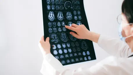
Imagine a scenario where a devastating condition like Parkinson’s disease could be identified long before its debilitating symptoms—tremors, stiffness, and balance issues—begin to disrupt daily life, offering a chance for early intervention. Parkinson’s disease (PD), a progressive neurodegenerative disorder, impacts millions globally by eroding dopamine-producing neurons in the brain, leading to both motor and non-motor challenges. By the time these symptoms become apparent, a staggering 60 to 80 percent of critical neurons may already be lost, making early detection not just beneficial, but potentially life-changing. Traditional diagnostic tools often fall short in catching the subtle brain alterations that mark the earliest stages of this condition. This gap has fueled a pressing need for innovative approaches that can peer deeper into the brain’s microstructure. Advanced imaging techniques, particularly Diffusion Tensor Imaging (DTI), are emerging as potential game-changers in this arena. As research continues to evolve, the promise of spotting PD before significant damage occurs offers hope for earlier interventions and improved patient outcomes. This article delves into how DTI is reshaping the understanding of PD, exploring its capabilities, recent findings, challenges, and the broader implications for care and treatment.
Understanding Parkinson’s Disease and the Need for Early Detection
The Impact of Parkinson’s Disease
Parkinson’s disease stands as the second most common neurodegenerative disorder after Alzheimer’s, affecting a vast number of individuals with its progressive decline in brain function. The condition primarily results from the loss of dopamine-producing neurons in the substantia nigra, a critical brain region. This depletion disrupts communication within the brain, manifesting in motor symptoms such as slowness of movement, muscle rigidity, and resting tremors, alongside non-motor issues like sleep disturbances and mood disorders. The insidious nature of Parkinson’s disease means that significant neuronal damage often precedes noticeable symptoms, with estimates suggesting that up to 80 percent of these vital cells may be gone by the time a diagnosis is made. This delayed recognition severely limits the window for interventions that could potentially slow the disease’s advance, underscoring the urgent demand for diagnostic tools capable of identifying changes at a much earlier stage.
Beyond the personal toll on patients, Parkinson’s Disease (PD) places a substantial burden on healthcare systems and families, as the disease progresses to impair daily functioning and independence. Early detection could revolutionize management strategies, offering a chance to implement therapies before irreversible damage sets in. The challenge lies in the subtlety of initial brain changes, which often evade detection through standard clinical assessments. Identifying these alterations could not only enhance the quality of life for those affected but also inform research into preventive measures. The focus on innovative technologies to bridge this diagnostic gap has never been more critical, as the aging population continues to grow, increasing the prevalence of such neurodegenerative conditions.
Limitations of Conventional Imaging
Conventional magnetic resonance imaging (MRI) has long been a cornerstone in evaluating brain health, capable of detecting gross structural changes like atrophy or iron accumulation in regions affected by Parkinson’s Disease (PD). However, its limitations become apparent when tasked with identifying the subtle microstructural damage that characterizes the early stages of the disease. Standard MRI often fails to capture the minute alterations in gray and white matter integrity that occur before significant atrophy or clinical symptoms emerge. This shortfall leaves clinicians reliant on observable symptoms for diagnosis, by which point substantial neuronal loss has already occurred, narrowing the scope for effective intervention.
The inadequacy of traditional imaging in early Parkinson’s Disease (PD) detection has driven the exploration of more sensitive methods that can reveal hidden brain changes. Without the ability to spot these early signs, patients may miss out on timely treatments that could slow disease progression. This gap highlights a critical need for advanced neuroimaging techniques that go beyond surface-level observations to probe deeper into the brain’s architecture. Such tools are essential for shifting the diagnostic timeline earlier, potentially transforming how PD is managed. As research pivots toward these innovative solutions, the hope is to equip medical professionals with the means to act proactively, addressing the disease before it fully manifests in debilitating ways.
What is Diffusion Tensor Imaging (DTI)?
Core Principles of DTI
Diffusion Tensor Imaging, commonly referred to as DTI, represents a specialized form of MRI technology that provides a unique window into the brain’s microstructural environment by measuring the movement of water molecules within tissues. Unlike standard MRI, which focuses on static anatomical details, DTI assesses the directionality and diffusion of water, offering insights into the integrity of both white and gray matter. This capability is crucial for detecting subtle disruptions in neural pathways that might not yet be visible through conventional imaging. A key metric derived from DTI is fractional anisotropy (FA), which quantifies the degree of tissue organization—lower FA values typically indicate damage or loss of structural coherence, while unexpected increases may point to other complex processes.
The significance of DTI lies in its ability to map out the brain’s intricate network of connections, revealing how well these pathways are functioning at a microscopic level. By generating detailed images of water diffusion patterns, DTI can highlight areas of compromise long before macroscopic changes, such as atrophy, become evident. This imaging approach is particularly valuable in neurodegenerative contexts, where early identification of tissue alterations could inform timely clinical decisions. As a non-invasive tool, DTI holds immense potential for enhancing diagnostic precision, providing a deeper understanding of brain health that goes beyond what traditional methods can achieve. Its application in research and clinical settings continues to expand, driven by the need for better tools to tackle elusive conditions.
Why DTI Matters for PD
In the context of Parkinson’s disease, DTI emerges as a promising avenue for detecting brain changes in regions critically affected during the early phases, such as the substantia nigra and corpus callosum. These areas often undergo microstructural alterations before any overt symptoms or visible atrophy appear, making them prime targets for early diagnostic efforts. DTI’s ability to capture these hidden changes offers a potential breakthrough, enabling clinicians to identify PD at a preclinical stage when interventions might be most effective. This early window could be pivotal in altering the disease trajectory, preserving neuronal function for as long as possible.
Moreover, the detailed insights provided by DTI into white and gray matter integrity could redefine how PD is approached diagnostically, offering a deeper look into the disease’s progression. By pinpointing specific regions of damage or dysfunction, this imaging technique allows for a more nuanced understanding of the disease’s impact on individual patients. Such precision is vital for developing targeted strategies that address the earliest signs of neurodegeneration. The potential of DTI to reveal abnormalities before they manifest clinically aligns with the broader goal of shifting PD management toward prevention and early action. As studies continue to validate its efficacy, DTI could become an indispensable tool in the fight against this challenging condition, offering hope for improved outcomes through earlier and more informed care.
Key Findings from Recent DTI Research in PD
Significant Brain Changes Detected
Recent research into the application of DTI in Parkinson’s disease has unveiled compelling evidence of its ability to detect microstructural brain changes that elude conventional imaging methods. One notable finding centers on the body of the corpus callosum, a major white matter tract, where significantly reduced fractional anisotropy (FA) values have been observed in PD patients compared to healthy individuals. This reduction suggests a loss of white matter integrity, potentially disrupting interhemispheric communication critical for coordinated movement and cognition. Studies have even proposed a specific FA cutoff value for this region as a diagnostic marker, demonstrating moderate sensitivity and specificity, which underscores DTI’s potential as a tool for identifying PD at earlier stages.
Equally intriguing are the unexpected increases in FA values reported in other brain areas, such as the right putamen and substantia nigra, among PD patients. These higher values might reflect compensatory mechanisms, inflammation, or gliosis rather than straightforward neuronal loss, painting a more complex picture of the disease’s impact on brain structure. Such variability in FA across different regions highlights that PD does not uniformly degrade tissue integrity but involves dynamic responses within the brain. These findings suggest that DTI can capture a spectrum of changes, offering a richer dataset for understanding neurodegeneration. As research builds on these observations, the nuanced data from DTI could refine diagnostic criteria, providing a clearer path toward early and accurate identification of PD.
Asymmetry in Brain Alterations
Another striking revelation from DTI studies is the asymmetry in brain changes among PD patients, often mirroring the side of the body where motor symptoms first manifest. Regions like the putamen and thalamus frequently show differing FA values between the right and left sides, with the affected side sometimes exhibiting higher or lower values that correlate with clinical presentation. This lateralization is significant, as it reflects the heterogeneous nature of PD progression, where one side of the brain may bear a greater burden of damage initially. Such findings suggest that DTI could map out these individualized patterns, offering a tailored view of how the disease unfolds in each patient.
The ability of Diffusion Tensor Imaging (DTI) to detect these asymmetrical alterations holds promise for personalizing treatment approaches. By identifying which brain regions are most impacted on a case-by-case basis, clinicians could design interventions—such as targeted therapies or deep brain stimulation—that address specific areas of concern. This correlation between imaging results and the side of symptom onset also strengthens the clinical relevance of DTI, as it ties directly to observable patient experiences. As further studies explore this asymmetry, the potential for DTI to guide precise, patient-specific care becomes increasingly evident, marking a shift toward more customized management of Parkinson’s Disease (PD). This aspect of DTI research opens new doors for understanding the disease’s variable impact across individuals.
Linking Imaging to Symptoms
DTI research has also established meaningful connections between imaging findings and the clinical severity of Parkinson’s disease, enhancing its practical utility. A notable observation is the inverse correlation between FA values in specific regions, such as the left putamen, and scores on clinical scales that measure motor and non-motor symptom severity. This relationship indicates that lower FA values in certain areas are associated with a greater burden of symptoms, providing a direct link between microstructural brain changes and real-world patient outcomes. Such correlations validate DTI as a tool that can reflect the current impact of PD on an individual’s health.
Interestingly, while FA values show ties to symptom severity, studies have often found no significant correlation with the duration of the disease. This suggests that the microstructural changes captured by DTI might not progress linearly over time but rather reflect the immediate state of brain integrity and its relation to symptom expression. This distinction is crucial for clinical applications, as it positions DTI as a snapshot of the current disease impact rather than a predictor of long-term progression. These insights pave the way for using DTI in routine assessments, offering objective data to complement subjective clinical evaluations. As this connection between imaging and symptoms is further explored, DTI could become a cornerstone in monitoring how PD affects patients at different stages.
Challenges and Future Potential of DTI in PD
Current Limitations
Despite the promising advancements of DTI in Parkinson’s disease research, several challenges remain that temper its immediate widespread adoption in clinical settings. One primary concern is the small sample sizes in many studies, which limit the generalizability of findings across the diverse spectrum of PD patients. These limited cohorts may not fully represent variations in disease severity, duration, or demographic factors, raising questions about the reliability of results when applied to broader populations. Additionally, technical issues such as motion artifacts during scans can introduce errors in DTI data, potentially skewing FA measurements and complicating interpretation.
Another hurdle lies in the complexity of interpreting FA values themselves, as changes can stem from multiple underlying causes, including neurodegeneration, inflammation, or compensatory processes, making it challenging to pinpoint the exact reason for the variations. This ambiguity makes it difficult to draw definitive conclusions from DTI scans without corroborating data from other sources. Furthermore, most current research employing DTI is cross-sectional, capturing only a moment in time rather than tracking changes over the course of the disease. This static approach restricts understanding of how microstructural alterations evolve, which is essential for assessing progression or treatment effects. Addressing these limitations will require concerted efforts to refine imaging protocols and expand study designs, ensuring that DTI’s potential is fully realized in practical applications.
Directions for Future Research
Looking ahead, the trajectory of DTI research in Parkinson’s disease points toward several key areas that could solidify its role in diagnosis and management. Expanding sample sizes in studies is a critical step, as larger, more diverse cohorts would provide a clearer picture of how DTI findings apply across different PD phenotypes and stages. Longitudinal studies, tracking patients over extended periods, are equally important to reveal how FA values and other DTI metrics change as the disease progresses. Such data could uncover patterns that predict clinical outcomes or responses to therapy, enhancing the predictive power of this imaging tool.
Integrating DTI with other neuroimaging modalities, such as functional MRI or positron emission tomography, offers a promising direction for advancing research. Combining structural insights from DTI with functional or molecular data could create a more comprehensive understanding of Parkinson’s disease (PD) pathology, bridging the gap between brain changes and their behavioral or symptomatic impacts. Additionally, refining the technical aspects of DTI—such as minimizing artifacts and standardizing measurement protocols—will be essential for ensuring consistency and accuracy in clinical use. As these research efforts advance over the coming years, from now through 2027 and beyond, DTI could transition from an experimental tool to a standard component of PD evaluation, driving forward the field of neurodegenerative diagnostics with each new discovery.
Broader Implications for PD Care
Transforming Diagnosis and Monitoring
The potential of Diffusion Tensor Imaging to transform the landscape of Parkinson’s disease diagnosis and monitoring is profound, particularly in its capacity to detect changes during the prodromal stage—before clinical symptoms fully emerge. By identifying microstructural alterations in key brain regions at an early point, DTI could enable diagnosis when interventions might have the greatest impact, potentially slowing disease progression and preserving quality of life. This shift toward preclinical detection aligns with the broader goal of proactive healthcare, where the focus is on prevention rather than reaction to advanced disease states. As a biomarker, DTI offers objective data that could complement traditional clinical assessments, reducing diagnostic uncertainty.
Beyond initial diagnosis, DTI holds value in tracking the course of Parkinson’s Disease (PD) over time, providing a means to evaluate whether treatments are effectively mitigating brain changes. This monitoring capability could inform adjustments to therapeutic plans, ensuring they remain aligned with a patient’s evolving needs. The ability to measure microstructural integrity repeatedly offers a dynamic view of disease progression, which is often lacking in current methodologies. As validation studies progress, incorporating DTI into routine clinical practice could redefine standards of care, equipping healthcare providers with a powerful tool to intervene earlier and monitor more precisely, ultimately enhancing patient outcomes in the face of this challenging condition.
Personalizing Treatment Approaches
DTI’s detailed mapping of brain changes in Parkinson’s disease opens up exciting possibilities for personalizing treatment strategies to match individual patient profiles. By highlighting specific regions of damage or asymmetry—such as in the putamen or thalamus—DTI can reveal unique patterns of neurodegeneration that vary from one person to another. This detailed insight allows clinicians to tailor interventions, whether through medication adjustments, physical therapy, or advanced options like deep brain stimulation, to target the most affected areas. Such customization could optimize therapeutic effectiveness, addressing the specific needs of each patient rather than relying on a one-size-fits-all approach.
Additionally, the role of DTI extends into the realm of clinical trials for new Parkinson’s Disease (PD) therapies, where it could serve as a precise endpoint for measuring treatment outcomes. By providing quantifiable data on brain changes, DTI enables researchers to assess the impact of experimental drugs or interventions with greater accuracy, potentially accelerating the development of effective solutions. This application could streamline the testing process, ensuring that promising treatments reach patients sooner. As DTI continues to gain traction, its integration into personalized care and research frameworks promises to enhance the precision of PD management, offering a future where treatments are as unique as the individuals they serve, driven by cutting-edge imaging insights.


