
The sheer volume of medical imaging studies performed daily has placed an unprecedented strain on radiologists, creating a critical bottleneck in the diagnostic process where early detection can mean the difference between life and death. With a global shortage of these highly trained specialists and an ever-increasing number of computed tomography (CT) scans being ordered, the risk of diagnostic delays and professional burnout is a growing concern for healthcare systems worldwide. This challenge has catalyzed the search for innovative solutions that can augment the capabilities of medical professionals without compromising the quality of care. The development of sophisticated artificial intelligence tools represents a promising frontier in addressing this issue, offering the potential to streamline workflows, enhance accuracy, and ultimately improve patient outcomes by ensuring that critical findings are identified more swiftly and reliably. The integration of such technology aims not to replace the expert radiologist but to empower them, turning a cascade of data into actionable insights with greater efficiency.
A New Ally in Medical Imaging
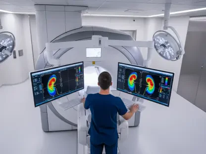
A groundbreaking collaborative effort between researchers at the University of Tartu, radiologists at Tartu University Hospital, and engineers has culminated in the creation of BMVision, an advanced artificial intelligence framework. This clinically validated tool is specifically engineered to analyze CT scans and assist medical professionals in identifying both malignant and benign kidney lesions with significantly improved speed and accuracy. The system functions as a powerful supportive instrument, acting as a second set of eyes for radiologists who are tasked with reviewing immense quantities of imaging data. Rather than operating as an autonomous diagnostic agent, BMVision enhances the diagnostic process by highlighting suspicious areas and providing precise measurements, thereby allowing doctors to focus their attention on the most complex aspects of a case. This human-in-the-loop approach leverages the strengths of both machine learning and human expertise, fostering a synergistic relationship that elevates the standard of care and addresses the operational pressures within modern radiology departments.
The utility of this AI solution extends profoundly into the realm of incidental findings, which are tumors discovered on scans performed for entirely unrelated medical reasons, such as evaluating abdominal pain or assessing injuries from trauma. A significant portion of kidney cancers are detected in this manner, making the thorough review of every scan a critical opportunity for early intervention. BMVision is designed to meticulously screen abdominal CT scans for subtle abnormalities that might otherwise be overlooked during a high-volume workday. By flagging these incidental lesions, the tool helps ensure that potentially life-threatening conditions are not missed, facilitating earlier diagnosis when treatment is most effective. This capability is central to its value proposition, as it directly contributes to improved patient prognoses by catching cancer at a nascent stage. The overarching consensus among its developers and early adopters is that the AI serves to amplify the radiologist’s ability to provide timely and precise diagnoses.
From Research to Real-World Results
The efficacy of the BMVision framework was rigorously substantiated in a retrospective study conducted at Tartu University Hospital, the results of which were published in the peer-reviewed journal Nature Communications Medicine. During the study, a team of six radiologists was tasked with reviewing 200 distinct CT scans, completing a full analysis for each case twice—once with the assistance of the AI tool and once without it. The findings were compelling, revealing that the use of BMVision reduced the time required to identify, measure, and formally report malignant kidney lesions by approximately one-third. This significant gain in efficiency did not come at the expense of accuracy; rather, it demonstrated how machine learning can streamline the most time-consuming aspects of image interpretation. The study provided robust evidence that modern AI tools have matured beyond the confines of research laboratories and are now capable of delivering a tangible, positive impact within the demanding environment of daily clinical practice, offering a validated solution to workflow inefficiencies.
Signaling its readiness for broader clinical adoption, BMVision has successfully obtained a CE marking, a certification that confirms its adherence to the rigorous health, safety, and environmental protection standards required for commercialization within the European Economic Area. This designation makes it the first commercially available AI tool specifically developed for the early and more accurate assessment of kidney cancer from CT imaging. Building on the success of its research phase, Tartu University Hospital is now in the process of fully integrating BMVision into its daily clinical workflow. The institution plans to have the system process all abdominal CT scans, a move anticipated to elevate the overall quality of diagnostics across the board. By systematically applying this technology, the hospital aims to facilitate earlier detection of kidney cancer on a large scale, which is expected to translate directly into improved treatment options and better long-term outcomes for patients.
Pioneering a New Standard in Radiology
The successful development and implementation of this AI-driven tool marked a significant milestone in diagnostic medicine. Through a rigorous validation process and subsequent regulatory approval, the framework demonstrated its capacity to meaningfully reduce the time radiologists spent on lesion analysis while simultaneously supporting their diagnostic accuracy. The integration of this technology into a major university hospital’s daily operations provided a clear and practical blueprint for how artificial intelligence could be thoughtfully woven into existing clinical pathways. This achievement not only provided a powerful solution to the mounting pressures faced by radiology departments but also established a new benchmark for the clinical utility of machine learning in oncology. The project served as a definitive proof of concept, showing that a collaborative approach between academia, clinical practice, and industry could yield innovations that directly enhanced patient care and operational efficiency.