
The global landscape of medical imaging is currently grappling with a paradoxical challenge where the demand for diagnostic services is skyrocketing while the traditional pathways for training the next generation of radiographers are becoming increasingly congested. As healthcare systems integrate more advanced modalities such as high-resolution Computed Tomography and rapid Magnetic Resonance Imaging into standard care pathways, the pressure to produce highly skilled practitioners has reached a critical juncture. Academic institutions and hospital departments are finding that the old model of “see one, do one, teach one” is no longer sufficient to meet the rigorous safety and efficiency standards required in modern medicine. This shift has necessitated a move toward simulation-based education, a transition that is not merely a technological trend but a fundamental restructuring of how clinical competency is defined and achieved. By creating a bridge between the controlled environment of the classroom and the unpredictable nature of the hospital ward, simulation offers a sustainable solution to the “clinical placement bottleneck” that has historically limited student enrollment.
The implementation of these high-fidelity environments allows for a more nuanced approach to learning, where students can engage in trial and error without the ethical or physical risks associated with live patient care. In a field where radiation protection and precision are paramount, the ability to fail safely is an invaluable pedagogical tool. These simulation suites do more than just replicate the layout of an X-ray room; they create a psychological space where learners can build confidence and technical mastery before ever touching a patient. As the industry moves toward 2027 and beyond, the integration of these tools is becoming a cornerstone of radiography curricula, ensuring that students are not just passive observers in a busy clinic but are instead active participants in their own skill acquisition. This evolution marks a significant move toward a more flexible and resilient model of medical education, one that prioritizes evidence-based competency over the mere accumulation of supervised hours in a high-pressure clinical setting.
Socio-Economic Pressures and the Training Gap
The primary driver for reforming radiography education is the widening gap between the soaring demand for diagnostic services and the available workforce capacity in modern healthcare systems. With imaging modalities like CT and MRI seeing annual growth rates exceeding 5%, the reliance on diagnostic data for everything from routine screenings to emergency interventions has never been higher. This surge is fueled by an expanding elderly population and more complex chronic health needs, leading to significant vacancy rates and an urgent requirement for new professionals who can hit the ground running. Despite the clear need for more staff, simply increasing student enrollment is frequently hampered by the physical and logistical limits of hospital imaging departments. Since traditional training mandates a specific number of supervised hours with live patients, the lack of clinical space and available mentors creates a significant barrier to graduation, leaving many potential students stuck in an educational limbo.
This bottleneck is further exacerbated by the increasing complexity of the equipment and the high turnover of patients in modern clinical environments. In a busy urban hospital, a student may find themselves relegated to the role of an observer because the pace of the department does not allow for the slow, methodical instruction required to master difficult positioning or software interfaces. Simulation provides a critical release valve for this pressure by decoupling academic progress from these physical and time-related constraints. By moving a portion of the technical training into university-based simulation labs, institutions can ensure that every student receives a standardized, high-quality education regardless of the patient volume at their assigned clinical site. This shift allows the healthcare system to expand its training capacity without overtaxing the already strained resources of front-line imaging departments, effectively creating a more scalable model for workforce development that can adapt to the needs of 2028 and beyond.
A Taxonomy of Modern Simulation Tools
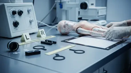
The variety of simulation technologies available to modern educators allows for a comprehensive and tiered approach to skill development that mirrors the complexity of the profession. Part-task trainers and anthropomorphic phantoms, such as the PIXY phantom, are essential for the foundational stages of learning, enabling students to practice precise positioning and exposure techniques without the risks associated with unnecessary radiation. These physical models provide a tactile experience that is essential for mastering the technical nuances of X-ray production, such as understanding how the angle of the tube affects anatomical distortion. Unlike practicing on a fellow student, which is ethically restricted due to radiation concerns, phantoms allow for the repeated execution of actual exposures, providing immediate feedback through the resulting images. This hands-on interaction with the hardware builds the muscle memory required for the high-stakes environment of a real trauma bay or orthopedic clinic.
Complementing these physical tools are digital innovations, including immersive Virtual Reality environments and sophisticated computer-based platforms like ScanLabMR. These systems offer a “safe-to-fail” digital twin of the clinical world where students can navigate virtual X-ray rooms, manipulate complex console parameters, and observe the immediate impact of their decisions on image quality and patient dose. While some early versions of these systems struggled to replicate the subtle tactile feedback of manual palpation, the latest haptic interfaces are beginning to bridge that gap, allowing learners to feel the “resistance” of bony landmarks through digital controllers. This level of immersion is particularly effective for teaching the physics of imaging, as students can visualize the X-ray beam and its interaction with matter in ways that are impossible in the physical world. By integrating these diverse tools, educators can create a multifaceted curriculum that addresses technical proficiency, spatial awareness, and the underlying scientific principles of the trade.
The Human Element: Communication and Emergency Response
Beyond the technical mastery of the equipment, modern simulation also addresses the critical human element of radiography through the use of simulated patients and high-fidelity mannequins. Professional actors or trained individuals are frequently employed to help students practice bedside manner, history taking, and the explanation of complex procedures to patients who may be anxious or in pain. This component of training is vital because the ability to communicate effectively can directly influence the quality of the diagnostic image; a patient who understands why they must remain still is much more likely to cooperate during a difficult procedure. These interactions allow students to develop their professional identity and emotional intelligence in a controlled setting where a mistake in communication does not lead to a genuine patient complaint or a missed clinical detail. It provides a platform to practice de-escalation techniques and the management of patients with diverse needs, including those with cognitive impairments or language barriers.
In addition to communication, high-fidelity mannequins that can simulate physiological crises, such as anaphylaxis or cardiac arrest, are used to prepare students for the rare but life-threatening emergencies that can occur in the imaging department. These advanced simulators can exhibit real-time changes in blood pressure, heart rate, and respiratory status, often in response to the administration of contrast media or other interventions. For a radiography student, experiencing these scenarios in a simulation lab is often the only way to develop the fast-acting reflexes and teamwork skills needed to support a medical emergency team. Training for these “low-frequency, high-stakes” events is nearly impossible in a traditional clinical placement, as students are usually—and rightly—sidelined during real emergencies to allow the experts to take over. Simulation ensures that when the student becomes a qualified professional, they possess the practical knowledge and calm demeanor necessary to manage a crisis effectively while maintaining the safety of the patient.
Navigating the Regulatory and Global Landscape
The acceptance of simulation as a legitimate and essential component of clinical training has gained significant momentum over the last few years, a change that was significantly accelerated by the necessity of adapting to global health disruptions. Professional bodies and regulatory agencies, which were once staunchly protective of traditional hospital-based hours, have begun to recognize that the quality of the learning experience is more important than the physical location where it occurs. For instance, influential organizations like the Society and College of Radiographers have established frameworks that allow for a designated number of simulated hours to count toward the total requirements for professional registration. This regulatory shift reflects a broader global trend toward academic flexibility and a recognition that simulation can offer a more consistent and measurable standard of competency than the often-variable experiences found in different hospital placements.
Internationally, there is an ongoing effort to harmonize these practices through evidence-based criteria that ensure simulated training is not just a placeholder for “time spent” but a rigorous pedagogical exercise. Organizations like the European Federation of Radiographer Societies are actively working to define what constitutes a high-quality simulation, emphasizing that these programs must be grounded in clear, objective learning outcomes and led by specially trained educators. This move toward competency-based assessment is helping to break down barriers between different healthcare jurisdictions, making the profession more mobile and allowing for a more standardized level of care globally. As we look toward the 2026 to 2030 period, it is expected that these international standards will continue to evolve, with an increasing emphasis on the use of data-driven metrics from simulation platforms to validate a student’s readiness for independent practice. This transition from a time-based model to one focused on proven ability is essential for maintaining public trust in the rapidly advancing field of medical imaging.
The Strategy of Supplemental Learning and Rare Scenarios
One of the most transformative aspects of simulation is its ability to expose students to “rare events” and complex pathologies that they might never encounter during their standard hospital rotations. Much like pilots who use flight simulators to master emergency landings or engine failures, radiography students can use these platforms to manage high-stakes scenarios such as severe multi-system trauma or complicated pediatric imaging. In a traditional clinical setting, a student’s exposure to such cases is entirely dependent on chance; some may see a wide variety of pathologies, while others might spend their entire placement performing routine chest X-rays and extremity exams. Simulation levels the playing field by ensuring that every graduate has demonstrated competence in handling the most challenging aspects of the job, regardless of where they were physically trained. This proactive approach to education creates a more versatile workforce capable of handling the unpredictability of modern emergency departments.
Moreover, the simulation environment enhances the learning process by intentionally removing the high-pressure, high-volume stress of a functioning hospital. In a live clinic, a student might feel an immense amount of pressure to work quickly to keep up with a long line of waiting patients, which often leads to “recipe-following” rather than a deep understanding of the underlying physics and anatomy. Simulation suites allow for a “deliberate practice” model where a student can stop, analyze an error, and repeat a task until it is perfected. This slower, more reflective pace fosters a deeper conceptual understanding of how equipment adjustments—such as changing the kVp or the focal spot size—actually impact the resulting image and the radiation dose to the patient. By mastering these principles in a quiet, supportive lab, students develop a level of technical fluency that allows them to focus more on patient care and clinical judgment once they return to the fast-paced world of the hospital.
Overcoming Barriers and Embracing a Hybrid Future
Despite the overwhelming evidence supporting its benefits, the widespread implementation of high-fidelity simulation faces several significant hurdles that require strategic planning and investment. The initial cost of acquiring state-of-the-art equipment, such as VR suites, high-fidelity mannequins, and advanced phantoms, can be prohibitive for many academic institutions. Furthermore, these systems require ongoing software subscriptions, hardware maintenance, and a dedicated staff of technical experts to ensure that the equipment remains operational and the scenarios are clinically accurate. There is also the challenge of faculty development; many veteran educators may be unfamiliar with the latest digital tools and require intensive training to effectively integrate simulation into their teaching. Overcoming these barriers requires a commitment from both the public and private sectors to fund the infrastructure necessary to train the next generation of healthcare workers in a way that meets the demands of a high-tech medical environment.
The path forward for radiography training is almost certainly a hybrid model that blends intensive, university-based simulation with targeted, high-intensity clinical placements. In this future, students will use simulation to master technical console operations, radiation safety protocols, and basic positioning before they ever step foot in a hospital. This “pre-validation” of skills means that their time in the clinical environment can be much more focused and productive, allowing them to concentrate on the nuances of patient interaction and the interpretation of complex clinical cases rather than struggling with the basics of the machinery. This transition from a traditional, time-based model to a performance-based one ensures that the radiographers of the future are not just technically proficient, but are also deeply compassionate and prepared for the complexities of modern medicine. By embracing these technological advancements, the industry can create a more robust and sustainable pipeline of talent, ensuring that the critical bottleneck of clinical training becomes a thing of the past.