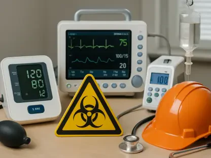

In the high-stakes environment of emergency medicine, trauma involving a pregnant patient presents an extraordinarily complex challenge, demanding rapid decisions that safeguard both maternal and fetal lives, while imaging emerges as a critical tool in guiding clinicians through the intricate balance of diagnosing injuries and minimizing risks to the unborn child. With trauma being the leading cause of non-obstetric mortality among pregnant women in the U.S.—often due to domestic violence, motor vehicle accidents, or falls—the urgency to act decisively cannot be overstated. Insights from experts like Dr. Margarita V. Revzin of Yale School of Medicine provide evidence-based direction for navigating these cases. This discussion delves into the essential principles for effective imaging, addressing common fears, optimal modalities, and the need for tailored approaches based on unique physiological factors. The goal is to equip clinicians with actionable strategies to manage these delicate situations with precision and confidence, ensuring the best possible outcomes for both mother and baby.
Maternal Stabilization as the Primary Focus
The bedrock of managing trauma in pregnancy lies in prioritizing maternal health, as it directly underpins the survival and well-being of the fetus. Severe injuries can precipitate maternal shock, a condition responsible for approximately 80% of fetal losses, with life-threatening trauma correlating to a staggering 40% to 50% fetal mortality rate. Stabilizing the mother—through immediate interventions to control bleeding, maintain blood pressure, and address critical injuries—must take precedence over all other considerations. Only after achieving this stability can attention shift to evaluating the fetus for signs of distress or injury. This sequential approach ensures that the foundation for fetal survival is secured first, acknowledging the profound interconnectedness of maternal and fetal outcomes in such dire circumstances. Clinicians must operate with this hierarchy in mind, recognizing that delays or misprioritization can have catastrophic consequences for both lives in their care.
Beyond initial stabilization, imaging becomes a vital step in assessing the full scope of maternal injuries and their potential impact on the pregnancy. Once the mother’s condition is under control, diagnostic tools can be employed to evaluate fetal viability, placental integrity, and any direct trauma to the unborn child. This dual assessment requires a nuanced understanding of how maternal injuries, such as internal bleeding or organ damage, might indirectly harm the fetus through reduced oxygen supply or other complications. The focus remains on ensuring that maternal recovery supports a safe environment for the fetus, while also identifying any immediate risks to the pregnancy that require intervention. This stage of care highlights the importance of interdisciplinary collaboration, where radiologists, obstetricians, and trauma specialists work in tandem to interpret imaging results and devise a comprehensive treatment plan tailored to the unique needs of both patients.
Overcoming Barriers of Radiation Concerns
A significant obstacle in trauma imaging for pregnant patients is the pervasive fear among clinicians of radiation exposure harming the fetus, often leading to hesitation in ordering necessary diagnostic tests. Contrary to common misconceptions, most imaging procedures, including computed tomography (CT) scans, deliver radiation doses well below thresholds considered harmful to fetal development. This misunderstanding can result in dangerous delays in diagnosis, potentially compromising timely treatment and endangering both mother and baby. Evidence underscores that the risk of not imaging when clinically indicated far outweighs the minimal risks associated with standard diagnostic radiation. Clinicians must be educated on these facts to make informed decisions, ensuring that fear does not impede critical care in emergency settings where every moment counts.
Addressing this issue requires a shift in perspective, supported by clear communication with patients and healthcare teams about the safety of imaging during pregnancy. Resources and guidelines from reputable organizations, such as those provided by the American College of Radiology, can help dispel myths and provide reassurance about the low risks involved. In stable patients, taking the time to discuss the necessity of imaging and the minimal impact of radiation can alleviate concerns and build trust. However, in emergencies with unstable patients, the priority must be on immediate action, proceeding with necessary scans without delay. This balanced approach ensures that clinical decisions are driven by evidence rather than unfounded apprehensions, ultimately protecting maternal and fetal health through prompt and accurate diagnosis of injuries that might otherwise go undetected.
Selecting Optimal Imaging Modalities
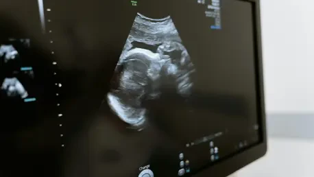
Choosing the right imaging modality is crucial in trauma cases involving pregnant patients, as each tool offers distinct advantages and limitations depending on the clinical context. Ultrasound, particularly through the Focused Assessment with Sonography for Trauma (FAST) protocol, stands as the preferred initial method due to its safety, lack of radiation, and widespread availability in emergency settings. It excels in quickly assessing fetal heart rate and basic uterine integrity in hemodynamically stable patients, providing immediate insights without posing risks to the fetus. However, ultrasound has notable shortcomings, missing up to 80% of placental abruptions—a leading cause of fetal death after maternal shock. Its inability to detect certain maternal or fetal skeletal injuries further limits its scope, often necessitating additional imaging to ensure a comprehensive evaluation of trauma severity.
When ultrasound falls short, other modalities like CT become indispensable, particularly in cases of severe abdominal or pelvic injuries where detailed visualization is critical. Despite involving ionizing radiation, CT is essential for unstable patients, offering rapid and precise detection of life-threatening conditions that might be missed by less sensitive tools. In emergencies, the need for speed often outweighs the minimal radiation risks, and scans may proceed without formal consent to avoid delays. For stable patients, informed discussions about radiation exposure, sometimes involving a medical physicist for fetal dose estimates, can guide decision-making. Additionally, the use of intravenous contrast in CT is considered safe during pregnancy and enhances injury detection, while overly conservative low-dose protocols may lead to repeat scans, inadvertently increasing exposure. This strategic selection of imaging tools ensures that diagnoses are both timely and accurate.
Navigating the Role of MRI and Contrast Use
Magnetic Resonance Imaging (MRI) offers a radiation-free alternative for trauma assessment in pregnancy, but its application is often constrained by practical limitations in acute settings. Due to longer acquisition times and limited availability in many emergency departments, MRI is less suitable for immediate trauma evaluations where speed is paramount. Instead, it finds its niche in subacute scenarios or when complex clinical questions arise, such as suspected neurological deficits or placental insufficiency requiring detailed imaging. The absence of ionizing radiation makes MRI an appealing option for in-depth assessments, but its role remains secondary to faster modalities like ultrasound and CT in the initial phases of trauma management. Clinicians must weigh these logistical factors against the diagnostic benefits when considering MRI as part of the care plan.
Another critical consideration is the use of contrast agents in imaging during pregnancy, particularly with CT and MRI. Intravenous iodinated contrast used in CT scans is widely regarded as safe and significantly improves the detection of internal injuries, providing clarity that can be vital for treatment decisions. In contrast, gadolinium-based agents for MRI are approached with greater caution, recommended only when the benefits clearly outweigh potential risks to the fetus. Overly conservative strategies, such as avoiding contrast or using ultra-low-dose protocols, can sometimes backfire by reducing diagnostic accuracy and necessitating additional scans, thus increasing overall exposure. A thoughtful approach, guided by clinical necessity and supported by evidence, ensures that contrast use enhances diagnostic precision without compromising safety for either the mother or the unborn child.
Tailoring Approaches to Gestational Dynamics
The stage of pregnancy profoundly influences imaging decisions and associated risks in trauma cases, requiring clinicians to adapt their strategies accordingly. As gestation progresses, the fetus becomes increasingly susceptible to direct pelvic trauma, particularly in later trimesters when its size and position offer less natural protection. Simultaneously, maternal physiological changes—such as increased blood volume and organ displacement—heighten vulnerability to abdominal injuries and complicate diagnostic assessments. These evolving dynamics necessitate a tailored approach, where imaging protocols are adjusted to account for both the changing anatomy of the mother and the developmental stage of the fetus. This adaptability is key to selecting the most effective and safest diagnostic tools at each point in the pregnancy.
Furthermore, understanding gestational age helps clinicians anticipate specific risks and prioritize certain evaluations over others during trauma management. Early in pregnancy, the focus might center on maternal injuries with less immediate concern for direct fetal trauma due to the smaller size and protective environment. In contrast, later stages demand heightened attention to potential fetal injuries and placental issues, as the risks of direct impact and abruption grow. Imaging choices, whether ultrasound for initial triage or CT for detailed assessment, must reflect these shifting priorities. This customized strategy ensures that diagnostic efforts address the most pressing concerns at each phase, balancing the need for rapid intervention with the imperative to minimize any potential harm to the developing child through careful modality selection.
Reflecting on Best Practices for Future Care
Looking back, the journey of refining trauma imaging protocols for pregnant patients revealed a critical need for evidence-based clarity and decisive action in emergency settings. The past emphasis on maternal stabilization as the foundation for fetal survival stood as a guiding light, ensuring that clinicians addressed life-threatening conditions without hesitation. Debunking radiation myths proved transformative, as it empowered healthcare providers to utilize necessary imaging tools like CT without undue delay. The nuanced roles of ultrasound, CT, and MRI became clearer over time, each finding its place in the diagnostic arsenal based on patient stability and injury severity. Moving forward, the focus should shift to enhancing interdisciplinary training, ensuring that teams are well-versed in gestational-specific risks and imaging safety. Developing accessible guidelines and fostering open communication with patients about diagnostic needs will further improve outcomes, paving the way for safer, more confident trauma care in pregnancy.