
A pivotal new study reveals a significant and concerning level of variability in how pediatric nuclear medicine images are interpreted, a finding that carries profound implications for the diagnosis and treatment of some of the most vulnerable patients. Research published in Pediatric Radiology by Hoeksema, Hayatghaibi, and Sharp systematically dissects the differences that arise between the initial reading of a scan and a subsequent review by another expert. This analysis moves beyond simply acknowledging that physicians may disagree; it delves into the fundamental reasons for these discrepancies, identifying factors ranging from individual analytical styles to the technology used in the process. The investigation uncovers a critical need for systemic change, proposing a multi-pronged approach involving standardization, enhanced collaboration, and the strategic integration of advanced technology to ensure that every child receives the most accurate diagnosis possible, thereby safeguarding their health and future.
Uncovering the Roots of Diagnostic Variability
The Central Challenge in a Specialized Field
The core revelation of the research is its direct challenge to the long-held assumption of uniform interpretation among highly trained medical professionals in the specialized field of pediatric nuclear medicine. This imaging modality is indispensable, providing unique functional and physiological data that other techniques like MRI or CT scans cannot capture, making it crucial for diagnosing complex conditions in children. However, the study demonstrates that the path from image acquisition to diagnostic conclusion is fraught with potential for significant divergence. This variability is not merely a matter of minor disagreement but can lead to fundamentally different clinical assessments. The findings serve as a critical wake-up call, suggesting that the existing framework for image interpretation may not be as robust or reliable as previously thought. It underscores a pressing, system-wide need to re-evaluate current practices, training protocols, and quality assurance measures to bridge the gap between expert opinions and achieve a higher standard of diagnostic consistency.
The research meticulously details how this interpretative variability is a notable concern even among seasoned physicians, a fact that complicates the diagnostic landscape for pediatric care. The complexity of nuclear medicine images, which depict physiological processes rather than just anatomical structures, inherently allows for a greater degree of subjective analysis. The study by Hoeksema and colleagues systematically explores this challenge, shifting the conversation from anecdotal evidence of disagreement to a data-driven analysis of a systemic issue. This core finding disrupts the perception of diagnostic imaging as a purely objective science and highlights the significant human element involved. It forces the medical community to confront uncomfortable questions about the reliability of current diagnostic pathways and emphasizes the urgent necessity for systemic improvements. The ultimate goal is to minimize the impact of individual subjectivity and ensure that every diagnostic report is grounded in a more standardized, evidence-based framework that prioritizes patient safety and optimal outcomes above all else.
Pinpointing the Sources of Discrepancy
A deep analysis of the cases examined in the study pinpointed three primary drivers behind the diagnostic inconsistencies, the first of which is the profound influence of individual interpretation styles. Each physician develops a unique method for analyzing images, a process shaped by their specific training, years of experience, and individual cognitive biases. This inherent subjectivity means that one expert might prioritize certain visual cues or data points over others, leading them down a different diagnostic path from a colleague viewing the exact same scan. Furthermore, the study highlights the critical role of contextual information. The clinical history, symptoms, and preliminary diagnoses provided alongside an imaging study can significantly frame or even bias an interpretation. Discrepancies frequently arose when primary and secondary reviewers were given different levels or types of this contextual data, demonstrating that a radiologist’s conclusion is not formed in a vacuum but is heavily influenced by the narrative accompanying the patient’s case.
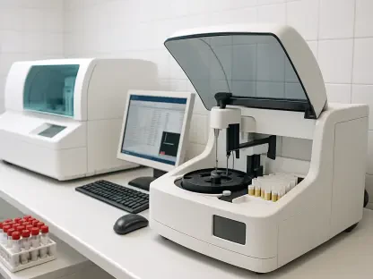
Beyond human factors, the research identified the technology itself as a significant source of variability. The specific software, analytical algorithms, and processing techniques used to render and analyze imaging data can yield slightly different quantitative and qualitative outputs. These subtle technological variations can, in turn, nudge a physician’s interpretation in a particular direction. For instance, one software package might calculate a particular metabolic rate differently than another, or a specific algorithm may highlight an area of concern that another does not. This points to a critical need for greater awareness among clinicians about the capabilities and limitations of their diagnostic tools, as well as a push toward greater standardization of the technology employed across different healthcare institutions. Without this uniformity, the field risks creating a system where a patient’s diagnosis could partly depend on the brand of software used at their hospital, a variable that has no place in high-stakes medical decision-making and must be addressed to ensure equitable care.
The High Stakes of Inconsistent Interpretations
Impact on Patient Care and Treatment Planning
The clinical consequences of these interpretative discrepancies are far-reaching and directly impact the well-being of young patients. One of the most striking revelations from the study is that divergent readings can precipitate completely different clinical recommendations, fundamentally altering a child’s entire treatment journey. For example, one interpretation might suggest a benign condition requiring only observation, while another reading of the same scan could indicate a malignancy necessitating immediate and aggressive intervention, such as surgery or chemotherapy. This level of inconsistency can lead to devastating outcomes, including critical delays in life-saving therapies, the initiation of inappropriate or unnecessarily invasive procedures, and immense confusion and anxiety for the clinical team and, most importantly, the patient’s family. The entire chain of care, from diagnosis to treatment and follow-up, is predicated on the accuracy of the initial imaging report, and when that foundation is unstable, the risk to the patient escalates dramatically.
This variability in diagnostic reporting creates a ripple effect of uncertainty that can undermine the trust between families and their healthcare providers. When parents receive conflicting information about their child’s condition, it can lead to a profound sense of distress and a loss of confidence in the medical system. They may be forced to seek third or even fourth opinions, navigating a complex and often intimidating healthcare landscape in search of a definitive answer. This process not only delays treatment but also places an enormous emotional and financial burden on the family. Furthermore, conflicting reports can create friction within the medical team itself, as different specialists may advocate for different courses of action based on disparate interpretations. This lack of consensus complicates collaborative care and can lead to operational paralysis, all while a child’s health hangs in the balance. Ultimately, the study makes it clear that diagnostic inconsistency is not just an academic concern but a critical patient safety issue that demands immediate and focused attention from the medical community.
Broader Systemic and Economic Effects
The impact of diagnostic discrepancies extends far beyond the individual patient, exerting significant pressure on the broader healthcare system. From an economic standpoint, conflicting interpretations are a major driver of inefficiency and wasted resources. When a primary and secondary reading do not align, it often triggers a cascade of additional medical procedures. This can include further consultations with other specialists, repeat imaging studies to clarify findings, or the ordering of more invasive and expensive diagnostic tests. Each of these steps adds to the overall cost of care, contributing to the already escalating healthcare expenditures. These are not just sunk costs but represent a misallocation of valuable resources that could be better used to treat other patients. The financial burden is borne by insurers, healthcare institutions, and ultimately, by the patients themselves, making diagnostic inconsistency a significant contributor to the economic strain felt across the entire healthcare ecosystem.
Beyond the direct financial costs, interpretative variability erodes the operational efficiency of healthcare delivery and raises fundamental questions about existing quality control measures. Inconsistent reports introduce delays into the clinical workflow, as care teams must pause to reconcile differences before a treatment plan can be finalized. This slows down the entire process of patient care, leading to longer hospital stays and inefficient use of clinical staff and facilities. Moreover, the study’s findings challenge the efficacy of current training paradigms and professional guidelines. If highly trained experts can arrive at such different conclusions, it suggests that there may be a fundamental need to re-examine how radiologists are trained, how they maintain their skills, and what reference standards they use in their practice. This calls for a systemic review of educational curricula, certification processes, and continuing medical education to foster a more consistent and reliable approach to pediatric imaging that can be trusted by patients and clinicians alike.
Forging a Path Toward Diagnostic Consensus
A Framework for Consistency and Collaboration
In response to the identified challenges, the study proposes a robust framework of solutions, with the development and adoption of standardized interpretation protocols at its core. Such protocols would establish a more uniform methodology for evaluating pediatric nuclear medicine studies, creating a clear, step-by-step framework that guides physicians through the analytical process. By outlining specific criteria for what to look for, how to measure it, and how to report it, these standards would reduce the impact of individual stylistic differences and cognitive biases. The goal is not to create a rigid, one-size-fits-all approach but to provide a consistent foundation that ensures all critical aspects of a scan are evaluated systematically across different practitioners and institutions. This move toward standardization would represent a major leap forward in diagnostic reliability, making the interpretation process more transparent, reproducible, and less susceptible to subjective variance.
Furthermore, the research places a strong emphasis on fostering a culture of enhanced collaboration among specialists as a powerful antidote to interpretative discrepancies. The study advocates for a multidisciplinary approach, where pediatric radiologists, nuclear medicine physicians, oncologists, and other relevant clinicians engage in regular, structured case review sessions. These collaborative forums create an environment where complex cases can be discussed openly, allowing professionals to share their unique perspectives, challenge each other’s assumptions, and build a consensus diagnosis. This process does more than just improve the accuracy of an immediate evaluation; it also serves as an invaluable tool for continuous professional development. By engaging in these peer-to-peer discussions, physicians can learn from one another, refine their interpretive skills, and stay abreast of the latest advancements in the field, ultimately elevating the collective expertise of the entire medical team and ensuring better outcomes for their patients.
Harnessing Technology for Enhanced Accuracy
Looking toward the future, the research highlights the transformative role that technology, particularly artificial intelligence (AI) and machine learning (ML), can play in refining diagnostic accuracy. These advanced systems hold immense promise as powerful assistive tools designed to support, not replace, the expertise of physicians. AI algorithms can be trained on vast datasets of medical images, enabling them to recognize subtle patterns, anomalies, and quantitative markers that may be imperceptible to the human eye. In a clinical setting, an AI tool could function as a vigilant “second reader,” automatically analyzing a scan and flagging areas of potential concern or highlighting inconsistencies between the image data and the preliminary interpretation. This would provide radiologists with a valuable safety net, helping to reduce the potential for human error, mitigate the impact of cognitive biases, and streamline the diagnostic workflow by focusing expert attention where it is most needed.
The successful integration of these advanced technological tools into routine clinical workflows represents a significant pathway toward achieving a more objective and reliable diagnostic standard in pediatric imaging. The implementation of AI and ML would require a concerted effort involving technology developers, healthcare institutions, and regulatory bodies to ensure the systems are safe, effective, and ethically deployed. This would involve rigorous validation of algorithms, the development of user-friendly interfaces that fit seamlessly into existing workflows, and ongoing training for clinicians to help them understand and leverage these new capabilities. By championing the thoughtful adoption of these innovations, the medical community can augment the irreplaceable judgment and experience of its human experts. This synergistic combination of human intellect and machine intelligence has the potential to build a more robust, transparent, and effective framework for interpreting pediatric nuclear medicine studies, ensuring that vulnerable young patients receive the most precise diagnoses possible to guide their care.