
Imagine a scenario where surgeons can peer into the molecular landscape of breast cancer during an operation, pinpointing malignant cells with unprecedented accuracy, a reality now unfolding through radionuclide imaging. This transformative technology enhances breast-conserving surgery (BCS), commonly known as lumpectomy, by focusing on removing cancerous tumors while preserving as much healthy breast tissue as possible, a delicate balance that hinges on achieving clear surgical margins—ensuring no cancer cells remain at the edges of the excised tissue. Historically, the challenge of positive margins has been significant, with 20-25% of patients requiring additional surgeries due to incomplete tumor removal. This not only burdens patients with added physical and emotional strain but also delays their recovery journey. Radionuclide imaging introduces a groundbreaking solution by using radiotracers to highlight cancer cells in real time, offering surgeons immediate feedback during the procedure. This innovation promises to reduce the need for follow-up surgeries, improve patient outcomes, and redefine precision in breast cancer treatment. The excitement surrounding this technology stems from its potential to bridge the gap between surgical intent and clinical success, providing a clearer path to complete tumor resection in a single operation. As this approach gains traction, it’s reshaping the landscape of breast cancer care, offering hope for more effective and less invasive interventions.
Tackling the Persistent Problem of Surgical Margins
The quest for clear surgical margins in breast-conserving surgery remains a critical focus for medical professionals aiming to minimize cancer recurrence. Positive margins—where cancer cells linger at the edges of removed tissue—pose a substantial risk, often necessitating a second operation to ensure all malignant tissue is excised. This issue affects a significant portion of patients, amplifying the urgency for more reliable intraoperative tools. Traditional methods, such as specimen radiography and frozen section analysis, have long been the standard for margin assessment. However, these techniques primarily rely on anatomical or visual cues, which can miss subtle signs of residual cancer, leading to uncertainty during surgery. The limitations of these approaches underscore the need for a paradigm shift toward more precise, biologically driven solutions that can offer definitive insights in the operating room.
Radionuclide imaging emerges as a compelling alternative by shifting the focus from structural to metabolic indicators of cancer. Unlike conventional tools, this technology detects the activity of cancer cells themselves, providing a deeper understanding of whether margins are truly clear. By offering real-time data during the procedure, it empowers surgeons to make informed decisions on the spot, potentially sparing patients the distress of additional surgeries. The impact of this shift is profound, as it addresses a long-standing gap in BCS by enhancing the likelihood of complete tumor removal in one session. This advancement not only improves clinical outcomes but also offers a psychological boost to patients, reducing the anxiety tied to uncertain surgical results.
Decoding the Mechanism of Radiotracer Technology
Central to radionuclide imaging is the use of a radiotracer known as 18F-Fluorodeoxyglucose (18F-FDG), a glucose analog that exploits the unique metabolic behavior of cancer cells. These cells, driven by the Warburg effect—a preference for aerobic glycolysis—absorb far more 18F-FDG than surrounding healthy tissue, becoming metabolically trapped due to their inability to fully process the compound. This differential uptake makes it possible to visualize malignant areas with striking clarity during imaging, providing surgeons with a molecular map of the tumor’s extent. The ability to highlight cancer at this level offers a significant edge over traditional methods, turning the operating room into a space for precision-driven decisions.
However, the effectiveness of 18F-FDG isn’t universal across all breast cancer types, presenting a nuanced challenge. Aggressive cancers, such as triple-negative variants, often exhibit high uptake, making them easily detectable. In contrast, less metabolically active tumors, like lobular carcinoma, may show weaker signals, complicating margin assessment. Additionally, timing plays a crucial role in optimizing results. With a half-life of roughly 110 minutes, 18F-FDG requires imaging within a narrow window of 1-3 hours post-injection to maximize the contrast between tumor and background tissue. These factors highlight the need for tailored approaches and precise coordination to fully harness the potential of radiotracer technology in surgical settings, ensuring its benefits are realized across diverse patient profiles.
Cutting-Edge Imaging Systems for Surgical Precision
The arsenal of radionuclide imaging includes several innovative systems designed to integrate seamlessly into the high-stakes environment of the operating room. Cerenkov Luminescence Imaging (CLI) stands out by capturing faint optical emissions from charged particles associated with radiotracers, using highly sensitive cameras to provide immediate visual feedback. Flexible Auto-Radiography (FAR) complements this by detecting beta particle interactions on a scintillating film, delivering exceptional spatial resolution to pinpoint radiotracer uptake in excised tissue. When combined, CLI and FAR create a synergistic effect, merging real-time imaging with detailed localization to enhance diagnostic accuracy. These tools are engineered for speed and practicality, ensuring they don’t disrupt the surgical flow while offering critical insights during procedures.
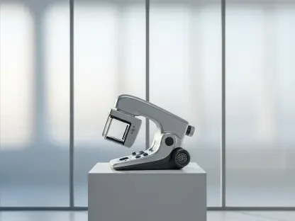
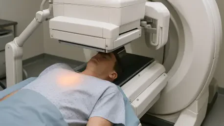
Beyond CLI and FAR, high-resolution Positron Emission Tomography/Computed Tomography (PET/CT) systems like the XEOS AURA 10 elevate precision to another level. These systems produce three-dimensional images with sub-millimeter detail, allowing for comprehensive margin assessment within minutes of tissue excision. Similarly, the LightPath® system employs both CLI and FAR to map radiotracer distribution, enabling surgeons to distinguish between malignant and benign areas swiftly. The portability and rapid imaging times—often under 10 minutes—of these devices make them viable for use even in standard hospital settings. Their design reflects a deep understanding of surgical needs, prioritizing ease of use alongside cutting-edge capabilities to support better decision-making and improve patient outcomes during BCS.
Transforming Patient Outcomes with Reduced Reoperations
One of the most compelling benefits of radionuclide imaging lies in its potential to significantly lower the rate of reoperations in breast-conserving surgery. Clinical studies have demonstrated remarkable effectiveness, with combined CLI-FAR approaches achieving specificity as high as 97.8% in identifying positive margins. This high specificity minimizes false positives, ensuring that surgeons avoid unnecessary removal of healthy tissue while still addressing cancerous areas. Sensitivity, though varying between 46.2% and 76.9% depending on the method, represents a substantial step forward compared to older techniques. These metrics translate into tangible improvements, offering a clearer path to successful single-procedure outcomes for many patients undergoing BCS.
Further evidence of impact comes from advanced systems like the XEOS AURA 10, which reports sensitivity and specificity rates of 91% and 94%, respectively, for prevalent cancers such as invasive ductal carcinoma. A striking statistic reveals that CLI-FAR has reduced reoperation rates by as much as 69% in certain studies, a figure that underscores the technology’s transformative potential. Another powerful insight is the number needed to treat: for every four patients managed with PET/CT systems, one additional surgery is avoided. These outcomes aren’t merely numbers on a page; they reflect real improvements in patient care, sparing individuals the physical burden and emotional weight of repeated interventions. The ripple effect of such advancements is a renewed sense of confidence in surgical approaches to breast cancer treatment.
Ensuring Safety and Practicality in Clinical Settings
Safety remains a paramount consideration in adopting any new medical technology, and radionuclide imaging excels in this regard for both patients and surgical teams. Radiation exposure to operating room staff during procedures is notably low, with median doses ranging from 15 to 38 microSieverts per surgery. This level is well below standard annual exposure limits, affirming the technology’s suitability for routine use without posing health risks to medical personnel. Such minimal exposure ensures that the benefits of enhanced margin assessment come without compromising the well-being of those delivering care, a critical factor in widespread acceptance of the technology.
Equally important is the practicality of integrating these imaging systems into diverse hospital environments. Devices like the XEOS AURA 10 are designed for portability, eliminating the need for specialized nuclear medicine facilities and making them accessible to a broader range of medical centers. Imaging processes are remarkably efficient, often completed in under 10 minutes, which prevents delays in surgical workflows and supports real-time decision-making. This adaptability allows even non-specialized hospitals to implement radionuclide imaging, democratizing access to cutting-edge care. By fitting seamlessly into existing protocols, these tools pave the way for broader adoption, ensuring that more patients can benefit from improved surgical precision regardless of where they receive treatment.
Addressing Barriers to Effective Implementation
Despite its promise, radionuclide imaging faces several hurdles that must be navigated to achieve its full potential in breast-conserving surgery. A notable limitation is the reduced visibility of tumors with low glucose metabolism, such as lobular carcinoma, which may not absorb 18F-FDG as avidly as more aggressive cancer types. This can result in missed detections, underscoring the need for complementary strategies or alternative radiotracers to address less metabolically active tumors. Additionally, larger specimens—those exceeding 4 centimeters—present challenges, as current imaging systems may struggle to capture comprehensive details across bigger tissue samples, potentially compromising accuracy in certain cases.
Another barrier lies in the logistical demands of the technology, particularly around timing and expertise. The optimal imaging window for 18F-FDG is between 60 and 120 minutes post-injection, requiring precise coordination within the often unpredictable pace of surgical schedules. Interpreting the resulting images also demands specialized training, necessitating close collaboration among surgeons, nuclear medicine specialists, and pathologists to ensure reliable results. False negatives remain a concern, especially for very small or multifocal tumors that may evade detection. Moreover, a learning curve exists for new users, which can initially slow adoption as teams build proficiency. While these challenges are significant, they are not insurmountable, pointing to areas where innovation and education can further refine the application of this powerful tool in clinical practice.
Charting the Future of Surgical Innovation
Looking ahead, the field of radionuclide imaging is poised for significant advancements that could solidify its role in breast cancer surgery. Efforts are underway to standardize protocols, such as defining consistent injection-to-imaging intervals and radiotracer dosages, to ensure uniformity and reliability across different clinical settings. Developing larger scintillators for Flexible Auto-Radiography is another promising direction, aimed at overcoming limitations in imaging bigger specimens and expanding the technology’s applicability. These technical refinements are crucial for addressing current gaps and enhancing the overall effectiveness of margin assessment during BCS.
Beyond hardware improvements, there’s a strong push toward enhancing diagnostic precision through better image interpretation algorithms. These advancements aim to minimize false negatives and boost confidence in identifying positive margins, even in complex cases. Multidisciplinary collaboration is also gaining emphasis, fostering partnerships among various medical specialties to create a more integrated approach to using radionuclide imaging. As training programs become more widely available, the barrier of expertise is likely to diminish, enabling more hospitals to adopt this technology. This momentum suggests a future where such imaging could become a standard component of breast cancer surgery, fundamentally improving outcomes and setting a new benchmark for precision in patient care.