
In the evolving landscape of cancer treatment, a groundbreaking approach is emerging that could transform how the effects of radiation on the human body are understood and managed, particularly in areas beyond the targeted tumor. This exploration delves into the subtle yet significant alterations in the femoral head and neck among prostate cancer (PCa) and rectal cancer (RCa) patients undergoing helical tomotherapy (HT), an advanced form of radiation therapy known for its precision. Through the innovative use of megavoltage computed tomography (MVCT) imaging, researchers have begun to uncover radiation-induced changes in bone tissue that are often invisible to traditional methods. These findings hold the promise of reshaping treatment monitoring, offering a pathway to detect early signs of bone stress before they escalate into serious complications. The focus on radiomics—a method of extracting detailed, quantitative data from medical images—brings a new lens to oncology, highlighting the potential for personalized adjustments in therapy to safeguard patient health while maintaining efficacy against cancer.
Unveiling the Power of Radiomic Analysis
Decoding the Science of Radiomics
Radiomics stands as a revolutionary tool in medical imaging, pulling intricate data from scans to reveal patterns and changes that escape the naked eye, particularly in the context of cancer therapy where subtle tissue damage can have profound consequences. This approach goes far beyond traditional imaging by quantifying features such as texture, density, and variability in bone structures like the femur during pelvic radiation treatments for PCa and RCa. By converting visual information into measurable metrics, radiomics provides deeper insight into how radiation impacts non-target tissues. This capability is crucial for identifying early indicators of bone stress or remodeling, which could lead to complications if left unchecked. The significance lies in its potential to inform clinical decisions, allowing for adjustments in treatment plans to enhance safety without compromising the fight against cancer. As a non-invasive method, it offers a window into the body’s response, paving the way for more tailored therapeutic strategies.
The integration of radiomics into oncology also addresses a critical need for precision in monitoring the side effects of radiation therapy on surrounding healthy tissues. In patients undergoing treatment for pelvic malignancies, the femur often bears unintended radiation exposure, risking long-term damage such as fractures or necrosis. Radiomics captures microarchitectural shifts in bone tissue through features derived from MVCT scans, providing data that can correlate with the level of radiation dose received. This quantitative approach enables clinicians to track changes over the course of treatment, offering a dynamic view of bone health that traditional imaging often misses. Furthermore, the method’s ability to analyze large datasets ensures that even minute variations are detected, supporting a more nuanced understanding of radiation’s impact. This level of detail is essential for developing interventions that protect bone integrity while maintaining the effectiveness of cancer treatment, marking radiomics as a cornerstone of modern oncology.
Synergy with Advanced Radiation Techniques
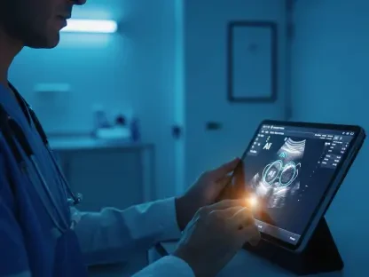
Helical tomotherapy, renowned for its ability to deliver highly targeted radiation doses, pairs seamlessly with radiomics by generating high-quality MVCT images that serve as the foundation for detailed analysis of bone changes. This precision technology minimizes exposure to healthy tissues, yet some radiation inevitably reaches areas like the femoral head and neck during pelvic cancer treatments. By leveraging MVCT data, radiomics tracks alterations in bone texture and density across multiple time points—before, during, and after therapy—offering a comprehensive view of how these tissues respond. This partnership between HT and radiomics fills a vital gap in current radiotherapy practices, where monitoring of non-target tissues often lacks the granularity needed for early intervention. The result is a powerful toolset that enhances the ability to safeguard patient health, ensuring that treatment remains both effective and as gentle as possible on surrounding structures.
Beyond mere detection, the combination of HT and radiomics facilitates real-time monitoring, a critical advancement in managing the unintended consequences of radiation therapy on bone health. The MVCT scans, typically used for patient positioning, are repurposed through radiomic analysis to assess subclinical changes in the femur, revealing patterns that might indicate early damage. This approach allows for a continuous feedback loop, where data on bone response can inform immediate adjustments to radiation doses or treatment fields. Such adaptability is particularly valuable for PCa and RCa patients, whose treatment plans often span weeks, increasing the risk of cumulative damage to nearby structures. By harnessing this synergy, the field of oncology moves closer to a model of care that prioritizes both tumor control and the preservation of quality of life, demonstrating how technology can bridge the gap between precision and patient safety in cancer therapy.
Insights into Bone Response and Clinical Impact
Effects of Radiation on Femoral Tissue
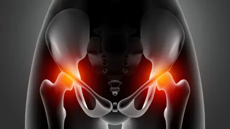
The impact of radiation on femoral bone tissue, as revealed through radiomic analysis, underscores the profound yet often hidden changes occurring at a micro level during cancer therapy for pelvic malignancies. Significant variations in radiomic features, particularly those related to tissue density and texture, were detected in the femoral head and neck of PCa and RCa patients undergoing HT. These shifts point to a process of bone remodeling triggered by radiation exposure, where the internal structure of the bone adapts in response to stress. Notably, a strong dose-dependent relationship emerged more clearly among PCa patients, with radiomic changes closely tied to the amount of radiation received. In contrast, RCa patients exhibited more variable responses, suggesting differences in tumor biology or treatment fields. These findings highlight the complexity of radiation’s effects on bone and the value of radiomics in capturing such nuances for better clinical understanding.
Further analysis reveals that specific radiomic features, such as those derived from intensity histograms and gray-level co-occurrence matrices, serve as sensitive indicators of bone alteration during therapy. These metrics reflect changes in density distribution and tissue heterogeneity, offering a detailed picture of how radiation reshapes the femoral microstructure over time. This level of insight is critical, as traditional imaging often fails to detect such early-stage modifications, which can precede more severe outcomes like fractures or pain. The ability to pinpoint these alterations also varies between anatomical regions, with the femoral head sometimes showing distinct patterns compared to the neck, likely due to differences in radiation exposure or bone composition. By mapping these responses, radiomics provides a foundation for identifying at-risk patients and tailoring interventions to mitigate long-term damage, ultimately enhancing the therapeutic balance between cancer control and tissue preservation.
Tracking Changes Over Treatment Phases
Monitoring radiomic features across different stages of treatment unveils distinct temporal patterns that align closely with radiation dose schedules, offering a potential early warning system for bone stress in cancer patients. Changes observed at mid-treatment and end-treatment phases in PCa and RCa patients undergoing HT were directly linked to the cumulative and fractional doses delivered, indicating that radiomics can capture the progressive impact of radiation on femoral tissue. This temporal sensitivity suggests that certain features could act as biomarkers, signaling bone health issues well before they manifest as clinical symptoms or visible damage on standard scans. Such early detection is invaluable, as it provides a window for clinicians to intervene—whether by adjusting radiation parameters or introducing supportive measures—potentially preventing complications that affect mobility or quality of life during and after therapy.
Delving deeper into these temporal dynamics, the ability of radiomics to track bone response over time also reveals variations between patient groups and treatment contexts, emphasizing the need for individualized monitoring approaches. For instance, the rate and nature of radiomic feature changes sometimes differed between PCa and RCa patients, possibly due to variations in radiation fields or underlying patient factors like age or bone density. These insights underscore the importance of longitudinal analysis, where data collected at baseline, midpoint, and conclusion of treatment paints a comprehensive picture of tissue evolution under radiation stress. By correlating these patterns with specific treatment milestones, radiomics not only enhances the understanding of bone response but also supports the development of predictive models. Such models could forecast which patients are most susceptible to adverse effects, guiding preemptive strategies that optimize outcomes in the challenging landscape of cancer care.
Shaping the Future of Personalized Oncology
Real-Time Adjustments for Safer Therapy
The integration of radiomic monitoring into cancer therapy via MVCT imaging presents a transformative opportunity to adjust radiation doses in real-time, protecting femoral bone health without undermining the effectiveness of treatment for PCa and RCa. By detecting subtle changes in bone tissue during HT, clinicians gain actionable insights that can prompt immediate modifications to treatment plans, such as reducing dose intensity in areas showing early signs of stress. This adaptability is a hallmark of personalized therapy, ensuring that each patient’s unique response to radiation is accounted for, rather than relying on standardized protocols that may overlook individual risks. The potential to minimize toxicity while maintaining tumor control marks a significant step forward, addressing one of the longstanding challenges in radiotherapy where balancing efficacy and safety remains paramount.
Moreover, real-time radiomic surveillance offers a practical framework for enhancing patient outcomes by preventing long-term complications that impact quality of life, such as bone pain or fractures. The ability to intervene mid-treatment based on MVCT-derived data shifts the focus from reactive to proactive care, where adjustments are made before damage becomes irreversible. This approach also fosters collaboration between radiation oncologists and medical physicists, who can use radiomic insights to refine dose distribution and shield vulnerable tissues more effectively. As a result, patients benefit from a treatment experience that prioritizes both their immediate cancer needs and their long-term well-being. The promise of such tailored interventions highlights how radiomics can redefine clinical workflows, embedding precision at every stage of therapy to ensure safer, more responsive cancer management.
Expanding Horizons for Radiomic Applications
Looking ahead, the findings from radiomic analysis of femoral bone changes lay a robust foundation for broader validation of these features as predictors of radiation-related complications in cancer therapy. Future research could focus on linking specific radiomic signatures to tangible clinical outcomes, such as fracture incidence, pain levels, or functional mobility, providing a clearer picture of their predictive power. Expanding studies to include larger, multi-center cohorts would strengthen the reliability of these metrics, ensuring they hold up across diverse patient populations and treatment settings. Additionally, correlating radiomic data with long-term follow-up results could help establish thresholds for intervention, guiding clinicians on when and how to act to prevent adverse effects. This direction promises to solidify radiomics as a trusted tool for risk stratification in oncology.
Equally compelling is the prospect of extending radiomic monitoring beyond the femur to other bony structures or cancer types exposed to radiation, potentially standardizing this approach across various therapeutic contexts. Investigating how radiomic features behave in different anatomical regions or under varying radiation modalities could uncover universal patterns of tissue response, broadening the applicability of this technology. Such efforts would also support the development of comprehensive guidelines for integrating radiomics into routine clinical practice, ensuring consistency in how bone health is assessed and managed during cancer treatment. By pushing these boundaries, the field moves closer to a future where radiomic surveillance becomes a cornerstone of radiation therapy, minimizing harm and maximizing safety for patients facing a wide array of malignancies, ultimately reshaping the landscape of personalized cancer care.