Physics World has published an article discussing the development and application of a portable multiphoton microscopy system aimed at rapid cancer diagnosis. This advanced imaging technology leverages femtosecond laser pulses for nonlinear optical imaging, producing high-resolution, pathology-quality images without sample processing or staining. The main objective is to enable real-time, in situ cancer detection, which could transform diagnostic procedures in clinical settings.
The Power of Multiphoton Microscopy
Advantages of Multiphoton Imaging
Multiphoton microscopy offers significant benefits over traditional confocal microscopy. By using femtosecond laser pulses to excite two- and three-photon processes, this technique produces high-resolution images without damaging biological tissues. The ability to visualize molecular and structural details in unprocessed samples holds considerable promise for rapid cancer diagnosis and personalized medicine.
Traditional confocal microscopy relies on linear optical processes, requiring extensive sample preparation, including slicing and staining to achieve contrast. In contrast, multiphoton microscopy employs nonlinear interactions where multiple photons simultaneously excite a fluorophore, resulting in higher spatial resolution and deeper tissue penetration. This innovative approach eliminates the need for stains and enables imaging of live tissues with minimal invasiveness.
The significance of multiphoton microscopy lies in its ability to enhance the speed and accuracy of diagnostic procedures. Its high-resolution imaging capability provides clinicians with detailed views of cellular structures and tissue architecture, facilitating better-informed decisions during medical consultations. Consequently, it holds promise for improving patient outcomes, streamlining workflows in pathology labs, and advancing personalized treatment strategies.
Role of Femtosecond Laser Pulses
Femtosecond laser pulses play a crucial role in achieving efficient multiphoton excitation. These ultrashort pulses, typically around 30 femtoseconds (fs), increase the likelihood of simultaneous photon interactions at the focal point, generating multiple nonlinear signals while maintaining low average power to avoid sample damage. The VALO Femtosecond Series lasers used in the system deliver pulses as short as 30 fs, enhancing the efficacy of two-photon and three-photon microscopy.
The usage of ultrafast femtosecond lasers is paramount in optimizing the multiphoton imaging process. Shorter pulse durations result in higher peak powers, essential for initiating multiphoton processes. However, maintaining low average power is critical to prevent potential heating or photodamage to the biological samples. This balance ensures that image quality is preserved, allowing clinicians to obtain reliable diagnostic information without compromising tissue integrity.
Advancements in femtosecond laser technology have enabled the development of compact and efficient imaging systems. The VALO Femtosecond Series lasers, renowned for their performance, extend the application range of multiphoton microscopy by delivering broad spectral bandwidths suitable for two-photon and three-photon excitation. This flexibility translates into enhanced imaging capabilities, providing scientists and medical professionals with powerful tools for a wide array of diagnostic and research purposes.
Flash Pathology’s Innovative Solution
Development of Portable Microscope
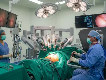
Flash Pathology, inspired by Marloes Groot’s work from Vrije Universiteit Amsterdam, has developed a compact, portable multiphoton microscope. With dimensions of 60 x 80 x 115 cm, this device is designed to provide rapid, on-site histologic feedback for excised tissues, focusing on usability and transportability.
The inception of the portable microscope by Flash Pathology aims to address the practical challenges posed by traditional pathology workflows. The compact design ensures that the device can be easily transported and utilized in various clinical settings, including hospitals, outpatient clinics, and remote locations. This portability enables real-time diagnostic feedback during surgical procedures, potentially reducing the reliance on centralized pathology labs and expediting the diagnostic process.
The integration of advanced optical components and user-friendly interfaces underscores the device’s practicality. Its innovative architecture allows clinicians to swiftly obtain high-resolution images of excised tissues, enabling immediate evaluation and decision-making. The device’s compact nature is particularly advantageous in surgeries where instant feedback on tissue margins is crucial, improving surgical outcomes and patient safety.
Clinical Application: Lung Cancer Diagnosis
One key application identified for this technology is lung cancer diagnosis. Lung biopsies are challenging because of difficulties in obtaining good diagnostic material from small lesions. Traditional histopathological analysis is time-consuming and requires extensive sample preparation. Multiphoton imaging can deliver immediate diagnostic feedback, reducing the time between biopsy and diagnosis.
In traditional lung biopsies, pathologists face significant challenges in acquiring sufficient diagnostic material due to the small size and diffuse nature of lesions. The conventional histopathological process involves extensive sample processing, staining, and examination under a microscope, which often spans several days. This delay can impede timely diagnosis and treatment, adversely impacting patient outcomes.
Multiphoton imaging addresses these issues by providing rapid and accurate visualizations of lung biopsy samples within minutes. Flash Pathology’s microscope, in recent studies, demonstrated the capability to image lung biopsy samples and provide feedback within six minutes post-excision, achieving an accuracy of 87%. This expedited diagnostic ability reduces the need for multiple biopsies during a single procedure, ultimately minimizing patient discomfort and enhancing clinical efficacy.
Technical Features and Performance
Generating Nonlinear Signals
The performance of the multiphoton imaging system depends on its ability to generate multiple nonlinear signals using a single ultrafast femtosecond laser. The system simultaneously produces second- and third-harmonic generation (SHG and THG), along with two- and three-photon fluorescence. These spectrally separated signals provide complementary diagnostic information essential for detailed tissue analysis.
Second-harmonic generation (SHG) and third-harmonic generation (THG) are pivotal in providing distinct diagnostic insights. SHG, highly responsive to non-centrosymmetric structures like collagen, is instrumental in assessing the structural integrity of tissues. THG, which arises at interfaces with differing refractive indices such as cell membranes, enhances cellular visualization. Combined, these signals furnish comprehensive details about tissue morphology, aiding pathologists in formulating more accurate diagnoses.
Two- and three-photon fluorescence, generated through femtosecond laser excitation, further add to the diagnostic prowess of multiphoton microscopy. These fluorescent signals provide contrast by highlighting specific molecular markers, revealing intricate details about cellular substructures and biological functions. This multiplicity in signal generation equips clinicians with a holistic view of tissue samples, ensuring thorough and precise diagnostic evaluations.
Demonstrated Accuracy
Flash Pathology’s microscope demonstrated its ability to image lung biopsy samples and provide feedback within six minutes post-excision, achieving an accuracy of 87%. This rapid diagnostic ability could significantly reduce the need for multiple biopsies during a single procedure, offering considerable benefits to patient care.
The validation of the device’s diagnostic accuracy underscores its potential for widespread clinical application. A notable aspect is the immediacy of feedback, which contrasts starkly with traditional methods. Real-time diagnostic information allows surgeons to make informed decisions without the need for prolonged waiting periods, thus improving patient management and expediting treatment plans.
This accuracy and speed are pivotal during surgical procedures, particularly in lung cancer biopsies where timely assessment of tissue margins is crucial. The ability to determine malignancy status swiftly can guide intraoperative decisions, potentially enhancing surgical outcomes by ensuring complete removal of cancerous tissues. Moreover, the reduced need for multiple biopsies minimizes patient discomfort and sampling errors, further solidifying the microscope’s role in augmenting clinical efficacy.
Future Applications and Enhancements
Widespread Clinical Use
Flash Pathology envisions the extensive clinical use of their multiphoton microscope, with applications across various fields requiring quick, intraoperative diagnostic feedback. Beyond lung cancer, the technology is being tested on other tissues in the Netherlands’ hospitals and research centers and for other cancer types such as mesothelioma in Glasgow.
The promising results from lung cancer applications have set the stage for broader clinical implementation. Multiphoton microscopy’s adaptability to different tissue types is a significant advantage, enabling its use in diagnosing various cancers and other pathological conditions. Hospitals and research centers across the Netherlands are conducting trials on different tissue samples, aiming to establish standardized protocols for multiphoton imaging in routine diagnostics.
One notable research effort involves testing the technology for mesothelioma diagnosis at the Queen Elizabeth Hospital in Glasgow. Mesothelioma, a challenging cancer often linked to asbestos exposure, requires precise diagnostic techniques due to its complex presentation. The rapid and detailed imaging offered by Flash Pathology’s microscope could facilitate earlier detection and accurate assessments of this aggressive cancer, potentially improving patient outcomes.
Integration with Artificial Intelligence
To further enhance their system, Flash Pathology plans to integrate artificial intelligence (AI) to aid in image interpretation. The goal is to develop a certified medical diagnostic device that not only generates images but also provides diagnostic conclusions, improving diagnostic accuracy and integrating seamlessly into clinical workflows.
Artificial intelligence (AI) integration represents a transformative advancement in the device’s capabilities. AI algorithms can analyze multiphoton images, identifying patterns and markers indicative of specific pathologies. This automated analysis enhances diagnostic accuracy by reducing human error and ensuring consistency in interpretations. Additionally, AI can expedite the diagnostic process by swiftly generating conclusions based on vast datasets and pre-programmed criteria.
Achieving certification for the AI-enhanced diagnostic device is a pivotal milestone. It signifies that the technology meets stringent medical standards, ensuring reliability and safety in clinical use. The integration of AI aligns with broader trends in healthcare digitization, where smart systems are increasingly employed to streamline workflows and enhance patient care. This confluence of cutting-edge imaging technology and AI-driven analytics promises to set new benchmarks in cancer diagnostics.
Conclusion
Physics World recently published an article about a breakthrough in cancer diagnosis with the development of a portable multiphoton microscopy system. This innovative imaging technology uses femtosecond laser pulses for nonlinear optical imaging, creating high-resolution images comparable to those seen in pathology labs. What is remarkable about this system is that it eliminates the need for sample processing or staining, which is often a time-consuming aspect of traditional cancer diagnostics.
The system’s main goal is to facilitate real-time, in situ cancer detection. This means that doctors and medical professionals would be able to diagnose cancer almost immediately during examinations, potentially transforming how cancer diagnoses are handled in clinical settings. The ability to achieve such rapid and precise diagnosis directly at the point of care could lead to earlier detection, better patient outcomes, and more efficient treatment planning. The development promises to be a game-changer, significantly expediting the diagnostic process and offering high-quality imaging capabilities previously restricted to more advanced, less accessible laboratory environments.