Sickle cell disease, a genetic condition characterized by abnormal hemoglobin, poses significant health challenges, particularly when it comes to bone complications like infarction and osteomyelitis. These issues, driven by the disease’s hallmark vaso-occlusive crises, often manifest with overlapping symptoms such as pain, swelling, and tenderness, making accurate diagnosis a critical yet daunting task. For many patients, especially children, distinguishing between these conditions early can mean the difference between timely treatment and prolonged suffering. Conventional imaging methods frequently fall short in providing clarity during the crucial early stages, leaving clinicians in need of more reliable tools. This article explores the transformative role of nuclear medicine, particularly dual-tracer bone scintigraphy, in addressing this diagnostic hurdle. By offering a non-invasive and precise approach, this technology is reshaping how medical professionals manage bone-related complications in sickle cell disease, ensuring better outcomes through targeted care.
Unraveling the Complexity of Sickle Cell Bone Challenges
Sickle cell disease disrupts normal blood flow as red blood cells take on a rigid, sickle shape, often leading to bone infarction—a condition where blocked blood supply causes bone tissue death, primarily in long bones like the femur and humerus. This results in severe pain and can significantly impair mobility, especially during acute crises. The risk doesn’t end there, as patients are also prone to osteomyelitis, a serious bone infection frequently caused by bacteria such as Salmonella. This heightened vulnerability stems from compromised immunity and tissue damage from repeated infarctions. Both conditions share strikingly similar clinical signs, including localized warmth, redness, and swelling, which complicates differentiation based solely on physical examination. Without precise diagnostic tools, clinicians face the risk of misdiagnosis, potentially delaying critical interventions. Understanding these overlapping presentations underscores the urgent need for advanced methods that can cut through the ambiguity and guide appropriate therapeutic strategies.
Beyond the immediate pain and discomfort, the long-term implications of these bone complications in sickle cell disease are profound, particularly for younger patients whose growth and development may be affected. Bone infarction can lead to chronic issues like joint damage if not managed properly, while untreated osteomyelitis risks systemic infection or permanent bone deformity. The challenge lies in the fact that early symptoms provide little to no distinguishing clues, often mimicking each other to a frustrating degree. Historical reliance on patient history and basic clinical assessments has proven insufficient, as these methods lack the specificity needed to pinpoint the underlying pathology. This diagnostic gray area not only heightens patient distress but also places a burden on healthcare systems striving to allocate resources effectively. As such, the medical community has turned to innovative technologies to bridge this gap, seeking solutions that offer clarity where traditional approaches falter, ensuring that each patient receives care tailored to their specific condition.
Shortcomings of Standard Diagnostic Imaging
When it comes to diagnosing bone issues in sickle cell disease, conventional imaging techniques like X-rays and ultrasounds often fall short, especially in the critical early stages. X-rays, typically the first line of imaging, require at least 10 days from symptom onset to reveal characteristic changes, missing the window for prompt intervention. Even then, the findings are often nonspecific, showing mere soft tissue inflammation without clearly indicating whether the issue is infarction or infection. This delay and lack of precision can lead to prolonged uncertainty, hampering the ability to start appropriate treatment swiftly. As a result, patients may endure unnecessary pain or face worsening conditions while clinicians await clearer diagnostic evidence, highlighting a significant limitation in managing acute bone crises effectively with these widely used tools.
Ultrasound, another common imaging method, offers some utility in detecting soft tissue abnormalities such as abscesses, but it struggles with deeper bone structures and fails to reliably distinguish between bone infarction and osteomyelitis. Its effectiveness is further limited by accessibility to certain anatomical areas, rendering it an incomplete solution for comprehensive diagnosis. This inadequacy is particularly problematic in pediatric cases, where minimizing diagnostic delays is essential to prevent long-term damage. The shortcomings of these traditional approaches often leave medical teams grappling with inconclusive results, forcing them to rely on empirical treatments that may not address the root cause. Such gaps in early detection emphasize the pressing need for more advanced diagnostic technologies that can provide definitive answers sooner, reducing the risk of missteps in patient care and paving the way for more targeted and timely medical responses.
Transformative Power of Nuclear Medicine
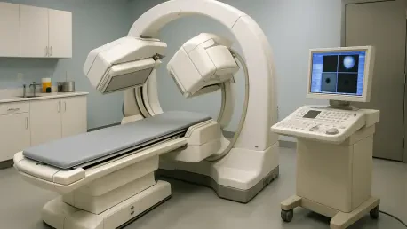
Nuclear medicine, through the use of dual-tracer bone scintigraphy, stands out as a revolutionary tool in differentiating bone infarction from osteomyelitis in sickle cell disease patients. This technique employs two distinct tracers—one to assess bone activity and another to evaluate bone marrow function—capturing images at various phases post-injection to analyze blood flow and metabolic changes. Unlike standard imaging, this method provides deeper insights into the physiological state of the bone, revealing patterns that are critical for accurate diagnosis. For instance, it can detect heightened activity in areas of concern, which might suggest either an infection or the revascularization phase of an infarction. By offering a non-invasive window into these processes, dual-tracer scintigraphy addresses the limitations of conventional methods, enabling clinicians to make informed decisions much earlier in the diagnostic journey.
A key strength of this approach lies in its ability to highlight distinct differences in bone marrow response, which is pivotal for distinguishing between the two conditions. In osteomyelitis, bone marrow scans typically show normal uptake, indicating preserved function despite infection, whereas bone infarction displays reduced uptake due to marrow necrosis. This differentiation proved invaluable in a documented case involving a young patient with sickle cell disease, where the technology identified both conditions in separate regions of the same bone. Such precision not only clarifies the diagnosis but also directly informs treatment strategies, ensuring that antibiotics are used for infections and supportive care is prioritized for infarctions. The adoption of nuclear medicine in these scenarios marks a significant advancement, reducing diagnostic ambiguity and enhancing the ability to tailor interventions to the specific needs of each patient, ultimately improving clinical outcomes.
Practical Insights from Clinical Applications
The real-world impact of nuclear medicine shines through in clinical scenarios involving sickle cell patients with ambiguous bone pain. Consider the experience of a 12-year-old boy who arrived at an emergency department with intense pain in his tibia, initially managed with pain relief that offered only temporary respite. As swelling worsened and lab tests suggested possible infection, standard imaging like X-rays and ultrasounds provided no definitive answers, showing only vague inflammatory changes. It was the application of dual-tracer bone scintigraphy that broke through the uncertainty, revealing osteomyelitis in one part of the bone and infarction in another. This clarity allowed medical teams to initiate targeted treatments—antibiotics for the infection and supportive measures for the infarction—demonstrating how this advanced tool can decisively alter the course of patient management in high-stakes situations.
Further illustrating its value, nuclear medicine minimizes the need for invasive procedures, a critical consideration especially for pediatric patients where reducing trauma is a priority. In the aforementioned case, the ability to non-invasively pinpoint the dual pathology avoided unnecessary exploratory measures that could have added to the patient’s distress. This diagnostic precision also helps in tracking the progression of bone conditions over time, as prior infarctions may show lingering changes in scans that need correlation with current symptoms. By integrating these findings with clinical history, healthcare providers can avoid misinterpreting old lesions as new issues, ensuring that each intervention is both timely and relevant. The practical application of this technology underscores its role as a cornerstone in modern diagnostics for complex conditions like sickle cell disease, offering a pathway to more confident and effective medical decision-making.
Shaping the Future of Patient Management
The integration of nuclear medicine into the diagnostic framework for sickle cell bone complications holds immense promise for enhancing patient care on a broader scale. By providing a reliable, non-invasive method to differentiate between conditions with starkly different treatment needs, it helps avoid the pitfalls of misdiagnosis, such as delayed antibiotics for infections or unnecessary procedures for infarctions. This precision is particularly impactful in pediatric populations, where early and accurate diagnosis can prevent long-term skeletal damage and improve quality of life. As awareness of dual-tracer scintigraphy’s benefits spreads among healthcare providers, its adoption could become a standard practice, ensuring that more patients benefit from timely and tailored interventions, reducing both morbidity and the burden on medical resources.
Looking ahead, the implications of this technology extend beyond individual cases to influence clinical protocols and research directions. Continued advancements in nuclear imaging techniques may further refine diagnostic accuracy, potentially lowering radiation exposure and expanding accessibility in diverse healthcare settings. The emphasis on correlating imaging results with comprehensive patient histories also sets a precedent for holistic care, encouraging multidisciplinary collaboration among hematologists, radiologists, and pediatric specialists. As the medical community builds on these foundations over the coming years, the focus remains on scaling up training and infrastructure to support widespread implementation. This forward-thinking approach not only addresses current diagnostic challenges but also lays the groundwork for innovative solutions, ensuring that patients with sickle cell disease receive the most effective care possible in an evolving medical landscape.


