
A significant portion of difficult-to-treat high blood pressure cases stems from a condition known as primary aldosteronism, where the adrenal glands produce excessive amounts of the hormone aldosterone. Identifying the precise source of this overproduction is critical, as it dictates a life-altering treatment path: surgery for a single, problematic gland or lifelong medication for issues affecting both. For decades, the definitive diagnostic tool has been adrenal venous sampling (AVS), an invasive and technically complex procedure that involves threading a catheter through the veins to collect blood directly from the adrenal glands. This method is not only uncomfortable for patients but is also available only at a limited number of specialized medical centers, creating a substantial bottleneck in care and leaving many patients without a clear diagnosis or optimal treatment plan. This diagnostic challenge has spurred a dedicated search for a more accessible, less invasive, and equally reliable alternative that could transform the management of this common form of secondary hypertension.
The Advent of a Non-Invasive Solution
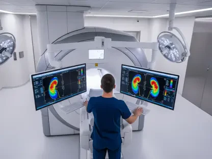
A Closer Look at Clinical Performance
Recent clinical data from a prospective Phase II study has offered a compelling glimpse into a potential replacement for the arduous AVS procedure. The investigation, centered on the radiodiagnostic agent [68Ga]Ga-Pentixafor, demonstrated notable success in distinguishing between unilateral and bilateral forms of primary aldosteronism. The results highlighted the agent’s high specificity, a crucial metric indicating its ability to accurately identify patients with the unilateral disease who would benefit from surgery. While its sensitivity was reported as moderate, the overall diagnostic performance has been hailed as a significant step forward. In the study, the PET/CT imaging procedure using [68Ga]Ga-Pentixafor was exceptionally well tolerated by participants, with no significant adverse events reported. This favorable safety profile, combined with the procedure’s non-invasive nature, presents a stark and welcome contrast to the risks and discomfort associated with the current gold-standard AVS, marking a pivotal moment in the quest for improved diagnostic protocols.
The patient experience has emerged as a particularly powerful aspect of the new imaging technique, with study findings revealing that nearly all participants expressed a strong preference for the [68Ga]Ga-Pentixafor scan over AVS. This overwhelming preference underscores the significant physical and psychological burden of the invasive catheterization process. The consistency of these results, which mirror findings from previous, smaller-scale studies, has provided a robust foundation of evidence. The reproducibility of the data across different research settings strengthens the case for this new method’s reliability and clinical utility. Buoyed by these positive and consistent outcomes, Pentixapharm Holding AG is now preparing to advance the radiodiagnostic agent into a large-scale Phase III clinical trial. This next stage will be critical in confirming the agent’s efficacy and safety in a broader patient population, potentially paving the way for regulatory approval and its eventual integration into standard clinical practice worldwide.
Understanding the Underlying Mechanism
The innovative power of [68Ga]Ga-Pentixafor lies in its ability to target a specific molecular marker, the chemokine receptor CXCR4. This receptor is found to be significantly overexpressed on the surface of aldosterone-producing adenomas, the small, benign tumors responsible for the unilateral form of primary aldosteronism. The radiodiagnostic agent is engineered by attaching a positron-emitting radioisotope, Gallium-68, to a molecule that selectively binds to the CXCR4 receptor. When introduced into the patient’s bloodstream, this compound circulates throughout the body and accumulates at sites with high concentrations of CXCR4—namely, the adrenal adenomas. A subsequent Positron Emission Tomography (PET) scan can then detect the radiation emitted by the Gallium-68, creating a detailed, three-dimensional image that precisely pinpoints the source of the aldosterone overproduction. This targeted approach provides functional and molecular information that goes far beyond the anatomical detail offered by traditional imaging like CT or MRI, enabling a far more precise diagnosis.
This molecular imaging strategy is a prime example of the growing field of theranostics, a paradigm that merges diagnostics and therapeutics. The principle is simple yet powerful: if you can see a target, you can treat it. The same CXCR4 receptor that allows [68Ga]Ga-Pentixafor to visualize the disease can also serve as a homing beacon for targeted therapy. This opens the door to a future where, upon confirming the presence of a unilateral adenoma via a Pentixafor PET scan, a physician could then administer a therapeutic version of the agent. This therapeutic counterpart would be labeled with a cancer-destroying radioligand, such as Lutetium-177, which would deliver a highly localized dose of radiation directly to the adenoma cells while sparing surrounding healthy tissue. This personalized, two-step approach promises to make treatment pathways more efficient, improve patient selection for surgical or medical interventions, and potentially offer new, non-surgical treatment options for this common condition.
Future Implications and Broader Impact
Redefining Patient Care Pathways
The potential integration of a non-invasive scan like the Pentixafor PET/CT into clinical workflows could fundamentally redefine the diagnostic and treatment pathway for millions of people with hypertension. By providing a simpler, safer, and more widely accessible alternative to AVS, this technology could empower general hospitals and smaller clinics to accurately subtype primary aldosteronism without the need for highly specialized interventional radiologists. This decentralization of diagnostic capability would dramatically reduce wait times, alleviate the travel burden for patients, and ensure that more individuals receive a definitive diagnosis sooner. Early and accurate identification of unilateral disease is paramount, as a timely adrenalectomy can cure hypertension and eliminate the long-term cardiovascular risks associated with aldosterone excess, such as stroke, heart attack, and kidney failure. This shift would not only enhance patient outcomes but also reduce the long-term economic burden on healthcare systems associated with managing chronic hypertension and its severe complications.
The introduction of this radiodiagnostic tool is also poised to have a ripple effect across multiple medical disciplines beyond endocrinology and cardiology. The CXCR4 receptor is implicated in a variety of other conditions, including numerous cancers and inflammatory diseases. The success of [68Ga]Ga-Pentixafor in the context of primary aldosteronism serves as a powerful proof of concept for its application in other areas. This validates the broader strategy of targeting the CXCR4 pathway for both imaging and therapy. As research continues, the theranostic platform built around this receptor could be adapted to diagnose, monitor, and treat a wide spectrum of diseases. This expansion represents a significant leap forward for personalized medicine, where treatments are tailored not just to a disease name but to the specific molecular fingerprint of an individual patient’s condition, heralding a more precise and effective era in modern medicine.
A New Horizon in Medical Imaging
The progress made with [68Ga]Ga-Pentixafor underscored a broader industry trend toward sophisticated radiopharmaceuticals that offer more than just anatomical snapshots. This evolution from structural to functional and molecular imaging provided clinicians with a deeper understanding of disease biology, which was crucial for making informed treatment decisions. The data from the Phase II trial not only highlighted a specific solution for an existing clinical problem but also reinforced the immense value of the theranostic approach. The ability to visualize a molecular target and then use that same target for therapy represented a paradigm shift that promised to streamline patient care and improve outcomes across various medical fields, setting a new standard for precision medicine. The successful trial and subsequent move toward Phase III development were seen as a significant validation of this innovative platform.