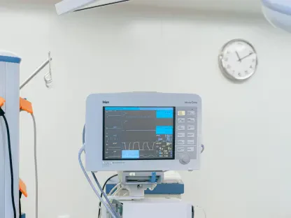

The sheer volume of chest radiography in modern medical facilities often places an immense cognitive burden on clinicians who must interpret millions of scans with absolute precision. With approximately 70 million chest X-rays performed annually in the United States alone, the potential for diagnostic fatigue or subtle oversight remains a persistent challenge for even the most experienced radiologists and emergency department physicians. This high-pressure environment necessitates a shift toward more integrated and intelligent diagnostic support systems that can operate at scale without compromising accuracy. Recently, the landscape of thoracic imaging has undergone a significant transformation following a major regulatory milestone achieved by a leading artificial intelligence developer. This advancement introduces a sophisticated layer of computer-assisted detection designed to harmonize with existing clinical workflows. By addressing the fundamental need for speed and reliability, this technology sets a new benchmark for how digital health tools are evaluated and deployed across the healthcare continuum during this era of rapid clinical evolution.
Enhancing Clinical Accuracy through Broad Diagnostic Integration
The Scope: Expanding Capabilities across Multiple Pathologies
The recent 510(k) Class II clearance of qXR-Detect by the FDA marks a pivotal expansion in the scope of computer-assisted detection for plain film chest X-rays. This software is specifically engineered to identify and localize critical findings across six distinct anatomical regions, including the lungs, pleura, heart/mediastinum/hila, bones, and even medical hardware. By providing such a comprehensive “6-in-1” solution, the technology effectively covers the vast majority of common abnormalities encountered in routine clinical practice. This broad utility ensures that practitioners do not have to toggle between multiple disparate tools to evaluate a single image, which significantly reduces the complexity of the diagnostic process. The integration of these six new indications brings the total number of FDA-cleared indications for the company’s portfolio to 26, solidifying its role as a primary driver of AI adoption in modern radiology departments and emergency rooms where every second of analysis counts.
Beyond the technical specifications of the detection algorithm, the software provides essential support to a wide range of medical professionals, including family medicine practitioners and emergency room staff. Because chest X-rays serve as the frontline of diagnostic imaging, these clinicians often encounter complex cases that require rapid triaging before a specialist radiologist can provide a final report. The ability of the AI to highlight and categorize positive findings allows for a more streamlined workflow, ensuring that the most urgent cases are prioritized effectively. This capability is not merely about identifying a pathology; it is about providing a structured layer of intelligence that supports clinical decision-making across the entire healthcare system. By automating the preliminary detection of findings in areas like the hila or the skeletal structure, the software acts as a secondary set of eyes that remains consistently vigilant, regardless of the patient volume or the time of day, thereby enhancing the overall safety net for patients.
Visual Clarity: The Role of Explainable Artificial Intelligence
A critical barrier to the adoption of artificial intelligence in healthcare has historically been the “black box” nature of many algorithms, where clinicians are presented with a result without a clear rationale. The qXR-Detect system addresses this challenge directly by prioritizing explainability through visual localization tools, such as bounding boxes and region-of-interest labels. Instead of providing a simple binary notification that an abnormality exists, the software visually identifies exactly where and why an alert was triggered on the X-ray. This transparency is vital for building trust between the technology and the clinician, as it allows the medical professional to verify the AI’s findings instantly against their own expertise. This collaborative approach ensures that the AI serves as an augmentative tool rather than a replacement, facilitating a more transparent diagnostic workup that benefits both the provider and the patient through clearer communication and more accurate results.
The practical implications of this explainable approach are particularly evident in the early recognition of subtle abnormalities, such as small lung nodules that might be obscured by surrounding structures. Experts at University Hospitals Cleveland Medical Center have noted that these tools are essential for identifying pathologies that could otherwise be overlooked during high-volume shifts or when clinicians are fatigued. By flagging these minute details early, the software provides a significant advantage in the management of chronic conditions and the early detection of life-threatening diseases. This proactive identification is a cornerstone of preventative medicine, as it allows for earlier intervention and better long-term patient outcomes. The focus on visual confirmation ensures that the software remains a practical tool for daily use, providing actionable insights that can be integrated into a patient’s medical record with high confidence, further bridging the gap between advanced data science and bedside clinical care.
Streamlining Regulatory Pathways and Future Deployment
Regulatory Innovation: The Predetermined Change Control Plan
The clearance of qXR-Detect is particularly noteworthy as it represents the first chest X-ray computer-assisted detection device to be approved with a Predetermined Change Control Plan (PCCP). This innovative regulatory framework is designed to accommodate the iterative nature of software development, allowing for updates and improvements to the algorithm without requiring a new 510(k) clearance for every architectural shift. In the past, the rigid nature of medical device regulations often meant that healthcare providers were stuck with older versions of technology while more advanced models sat in regulatory limbo. The implementation of a PCCP ensures that U.S. healthcare providers can benefit from the most current data models and technological refinements in real-time. This approach not only fosters a culture of continuous improvement within the AI sector but also ensures that the tools used in clinical settings are always operating at the highest possible level of performance as the underlying technology evolves from 2026 and beyond.
Furthermore, this regulatory milestone provides a scalable blueprint for how other medical AI solutions might be managed in the future. By pre-defining the scope of future modifications, the manufacturer demonstrates a high level of rigor and transparency that satisfies the stringent requirements of the FDA while maintaining the agility needed for modern software deployment. This balance between safety and innovation is crucial for the long-term sustainability of digital health solutions in the United States. Healthcare systems can now invest in AI platforms with the confidence that their technology will not become obsolete shortly after implementation. Instead, they are adopting a living system that adapts to new data and improved methodologies, providing a more consistent and reliable diagnostic service. This shift in regulatory strategy highlights the maturing relationship between technology developers and government oversight agencies, paving the way for more rapid advancements in the field of diagnostic radiology.
Global Standards: Validating Performance through Clinical Evidence
The path to FDA clearance was underpinned by extensive performance testing, including a multi-reader multi-case clinical study that demonstrated the algorithm’s consistency and reliability. These studies compared the AI’s performance against established success criteria, proving that it could meet or exceed the diagnostic accuracy of traditional methods. This commitment to robust clinical validation is essential for the global expansion of medical AI, which is currently utilized in 107 countries. The data gathered during these trials provided the necessary evidence to support the use of AI in high-stakes clinical environments, ensuring that the software performs reliably across diverse patient populations and different imaging hardware. Such rigorous testing protocols reinforce the credibility of the technology, making it a viable option for large-scale healthcare networks that demand high standards of evidence-based medicine before adopting new diagnostic tools into their standard protocols.
Health systems moved toward a more integrated model of care by incorporating these validated AI tools into their daily radiological workflows. To maximize the benefits of this technology, hospital administrators coordinated with IT departments to ensure seamless interoperability between the AI software and existing Picture Archiving and Communication Systems. Clinicians also underwent specialized training to interpret the localized findings effectively, ensuring that the visual cues provided by the software were translated into actionable patient care plans. Moving forward, institutions should continue to monitor the impact of these tools on diagnostic turnaround times and patient outcomes to quantify the return on investment. The successful deployment of this technology demonstrated that when AI is supported by strong clinical evidence and transparent regulatory frameworks, it became an indispensable asset in the fight against diagnostic error and a catalyst for improving the efficiency of thoracic imaging on a global scale.