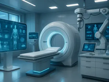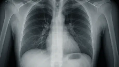

Imagine a world where diagnosing critical lung conditions like pulmonary embolism or chronic obstructive pulmonary disease (COPD) no longer requires exposure to radioactive tracers or lengthy, multi-step procedures, and this vision is becoming a reality thanks to a pioneering advancement in respiratory diagnostics. An Australian-based company, listed on the ASX under the ticker 4DX, has recently achieved a groundbreaking milestone with the FDA clearance of its CT:VQ imaging software. This technology marks a significant departure from traditional nuclear medicine scans by offering a non-contrast, CT-based ventilation-perfusion tool that evaluates airflow and blood flow in the lungs with unprecedented efficiency. By leveraging existing CT infrastructure, this innovation promises to transform how lung imaging is conducted, making it safer, faster, and more accessible to patients across diverse healthcare settings. The impact of this development could reshape standards of care in pulmonary diagnostics.
Revolutionizing Diagnostic Efficiency and Safety
The advent of CT:VQ imaging software introduces a transformative approach to lung diagnostics, addressing longstanding challenges associated with conventional ventilation-perfusion scans. Traditional methods often require two separate procedures using radioactive tracers, a process that can take over an hour and poses risks due to radiation exposure. In contrast, CT:VQ utilizes existing CT scanners to generate detailed ventilation and perfusion maps without additional equipment or contrast agents. This streamlining not only cuts down diagnostic time significantly but also eliminates the health risks tied to radioactive materials. Hospitals benefit from reduced scheduling burdens, as the technology integrates seamlessly into current workflows. For patients, this means quicker access to high-quality, quantitative images that aid in diagnosing serious conditions such as asthma or pulmonary embolism, all while prioritizing safety and comfort in a way that older methods could not match.
Beyond the immediate benefits of speed and safety, CT:VQ stands out for its ability to deliver superior diagnostic precision without compromising on accessibility. The technology has undergone rigorous clinical testing, showing strong agreement with single photon emission computed tomography (SPECT), the current benchmark in nuclear ventilation-perfusion imaging. Physicians have highlighted that the images produced are clearer, avoiding common pitfalls like tracer clumping or leakage that can obscure results in traditional scans. This clarity enhances diagnostic accuracy, providing clinicians with reliable data to make informed decisions about patient care. Moreover, the elimination of radioactive tracers aligns with broader healthcare goals of minimizing patient risk while maintaining or even improving the quality of diagnostic outcomes. As a result, this innovation sets a new precedent for how lung function can be assessed in clinical environments.
Expanding Access to Advanced Lung Imaging
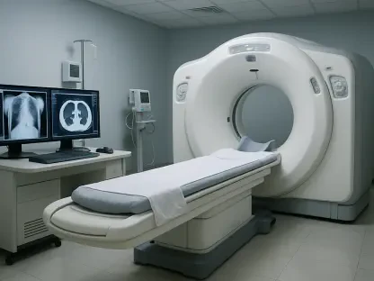
One of the most compelling aspects of CT:VQ technology is its potential to democratize advanced lung imaging across the United States. With approximately 14,500 CT scanners already in operation nationwide, this software can be deployed in a wide range of facilities, including smaller or regional hospitals that often lack specialized nuclear medicine departments. Previously, functional lung imaging was a resource-intensive process, largely confined to larger, well-equipped medical centers. By leveraging existing infrastructure, CT:VQ breaks down these barriers, ensuring that patients in underserved areas can access cutting-edge diagnostics without needing to travel to urban hubs. This widespread compatibility not only enhances equity in healthcare delivery but also positions the technology for rapid adoption in diverse clinical settings, fundamentally changing who can benefit from such advanced tools.
Additionally, the integration of CT:VQ into everyday medical practice is poised to reshape how frequently and proactively lung imaging is utilized. The removal of logistical hurdles, such as the handling and disposal of radioactive tracers, makes the procedure more feasible for routine use in managing chronic respiratory conditions or even as part of regular screenings. This could lead to earlier detection of issues like COPD, improving patient outcomes through timely intervention. The technology’s straightforward implementation into existing CT systems further reduces the learning curve for medical staff, facilitating smoother transitions in hospital workflows. As access to this diagnostic tool expands, it could encourage a shift toward more preventive approaches in respiratory care, addressing conditions before they escalate and reducing the overall burden on healthcare systems.
Commercial Potential and Market Impact
From a commercial standpoint, the FDA clearance of CT:VQ opens up a substantial market opportunity, particularly in the US, where the annual market for ventilation-perfusion scans is valued at over $1.1 billion. With more than one million such procedures performed each year, the potential for market penetration is significant. Established reimbursement rates and the ease of integrating this software into current CT systems provide a clear pathway for rapid rollout. By offering a safer and more efficient alternative to nuclear scans, there is a realistic prospect of displacing outdated methods entirely. Furthermore, the reduced risks and simplified process could expand the market itself, encouraging more frequent use in both acute and chronic disease management, thus driving demand beyond current levels and reshaping the economic landscape of pulmonary diagnostics.
Financially, the company behind CT:VQ is demonstrating robust growth and stability to support its ambitious expansion plans. Operating revenue for the full year ending June 30 has seen a remarkable increase of 56%, reaching A$5.9 million, driven by a growing Software-as-a-Service model. Nearly 195,000 scans were performed during this period, with quarterly volumes doubling compared to prior figures, signaling strong demand across domestic and international markets. Strategic cost reductions have trimmed annual expenses by A$6.5 million, while a significant A$10 million investment from a major player in medical imaging has bolstered liquidity. With substantial cash reserves and anticipated R&D tax credits, the financial foundation is solid, enabling focused efforts on capturing market share and potentially redefining industry standards over the coming years through sustained innovation and outreach.
Reflecting on a Milestone in Respiratory Care
Looking back, the FDA clearance of CT:VQ marked a turning point in the field of respiratory diagnostics, setting a new benchmark for safety and efficiency. This technology redefined patient care by offering a non-invasive, rapid alternative to nuclear scans, ensuring that conditions like pulmonary embolism and asthma were diagnosed with greater precision and less risk. Its integration with existing CT infrastructure broadened access, bringing advanced imaging to diverse healthcare settings and supporting equity in medical services. Supported by strong clinical validations and early adoption by leading institutions, this breakthrough captured significant attention and trust within the medical community. Moving forward, the focus should shift to scaling adoption, educating healthcare providers on the benefits, and exploring additional applications for CT:VQ in preventive care. This milestone serves as a reminder of how innovation can address critical gaps, paving the way for future advancements that continue to prioritize patient well-being and operational excellence.