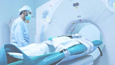
Nuclear medicine represents a transformative advancement in medical diagnostics, crucial for its ability to not only show the structure of our inner workings but also to reveal the function of our organs and tissues with great specificity. The practice harnesses the power of radiopharmaceuticals – radioactive substances – to shed light on various diseases, including cardiac conditions, cancers, and brain ailments.Its most significant application lies in its meticulous ability to pinpoint diseases, monitor organ function, and assist in plan formulation for patient care. Due to the informative nature of the physiological data it provides, nuclear medicine has risen to a critical position in the diagnostic realm. With the capacity to detect abnormalities early on in their development, this specialized field of medicine substantially aids in early intervention, treatment strategies, and better patient outcomes.Therefore, as technology progresses, nuclear medicine continues to play a key role, evolving the landscape of health diagnosis and therapeutics. This synergy of technology and medicine is pivotal in advancing healthcare, improving not just life expectancy but also the quality of life for patients globally.
The Science Behind Nuclear Medicine
Understanding Radioactive Tracers
Radiopharmaceuticals are integral to nuclear medicine and are administrated to patients in various ways – via injection, ingestion, or inhalation. These radioactive substances are engineered to target specific body parts such as organs, bones, or tissue, thanks to their unique chemical properties. Once inside the body, these tracers travel to their destined area and begin to emit gamma rays as they decay. This emission provides a source of energy that specialized imaging devices can detect.Through this process, healthcare professionals obtain not just static images but dynamic visualizations of how a patient’s body is functioning. This real-time insight allows for a closer examination of physiological processes, enabling the identification of disease-related irregularities that might not be visible through traditional imaging techniques. By combining structure with function, radiopharmaceuticals provide a more comprehensive understanding of a patient’s health, aiding in the precise diagnosis and management of various conditions. This functionality offers a stark contrast to other imaging methods that are limited to displaying only the structural aspects of the body.
Specialized Imaging Equipment
In the field of nuclear medicine, advanced imaging technology is essential for detecting illnesses at their inception. Key tools like gamma cameras, SPECT, and PET are used to trace radioactive markers in the body, showcasing physiological shifts indicative of diseases before any physical manifestations. Gamma cameras provide flat images, whereas SPECT and PET yield volumetric visuals that unveil the inner workings of the body in great detail.These imaging modalities are even more powerful when used in conjunction with CT or MRI scans, as they fuse anatomical detail with functional insight. This synergy allows for a more holistic analysis, facilitating the early discovery and treatment of medical conditions. The fusion of structural and functional imaging enables medical professionals to diagnose with greater accuracy and tailor treatments that are more targeted and effective.Collectively, these diagnostic machines are pivotal in nuclear medicine, providing a window into the body’s subtle molecular activities. Their ability to pinpoint the locations and degrees of pathological changes significantly boosts the capacity for early intervention, ultimately contributing to better patient outcomes.
Diagnostic Applications of Nuclear Medicine
Cardiac Function and Heart Diseases
Nuclear medicine plays a crucial role in diagnosing heart diseases by enabling the observation of cardiac function during activity. Through the evaluation of blood flow and the condition of cardiac muscles, nuclear cardiologists are equipped to identify a range of heart-related issues, such as coronary artery disease, heart failure, and variations in the heart’s size or shape. This technique goes beyond the capabilities of conventional imaging methods by providing more detailed insights, which is paramount for the early detection and tailored management of heart conditions. As a result, nuclear medicine is a key contributor to enhancing patient care, offering the potential for life-saving early treatments and more effective, individualized management plans for those with heart conditions. The advanced insights garnered from this approach underscore its significance in the realm of cardiac healthcare, emphasizing its contribution to better patient prognosis and health outcomes.
Detecting Cancer and Brain Disorders
Positron Emission Tomography (PET) scans are vital in oncology, playing a crucial role in accurately diagnosing and determining the extent of cancer. These scans are adept at pinpointing the exact location of tumors by tracking cell activity levels, thus distinguishing between benign and malignant masses with precision. Their efficiency extends to directing biopsies, shaping treatment strategies, and evaluating the effectiveness of cancer therapies over time.Beyond cancer diagnostics, the significance of PET scans in neurology is undeniable. They are key in delving into the complexities of Alzheimer’s disease and other forms of dementia. By observing cerebral blood flow and metabolic patterns, PET scans contribute to discerning between various dementia disorders. This is instrumental in tailoring patient care to specific needs.The integration of PET scans into medical diagnostics has revolutionized our approach to disease management, elevating the precision of treatment across a spectrum of conditions. Their non-invasive nature and ability to provide real-time insights into cellular function make them an indispensable tool in modern medicine. Whether it’s navigating the challenges posed by cancer or demystifying the intricacies of brain diseases, PET technology stands at the forefront, delivering critical information that empowers healthcare professionals to make informed decisions and offer targeted interventions.
Assessing Other Organ Systems
Nuclear medicine extends its diagnostic reach well beyond heart and brain evaluations, serving as a powerful tool for assessing various organ systems. For instance, lung function can be meticulously reviewed using imaging techniques that highlight both the ventilation and blood flow, which is vital for spotting conditions such as pulmonary embolisms or chronic obstructive pulmonary disease.When it comes to the kidneys, nuclear medicine steps in to gauge functionality and discover potential blockages through renal scans. This is pivotal for patients presenting symptoms that may indicate kidney disorders. Furthermore, for those experiencing gallbladder issues, hepatobiliary imaging offers insights into the organ’s condition, allowing for accurate diagnosis and subsequent treatment planning.Thyroid health is another area where nuclear medicine proves invaluable. By conducting thyroid scans, physicians can pinpoint whether a thyroid gland is overactive or underactive. This information is critical, as the thyroid plays a major role in metabolism and overall bodily function. Through these scans, clinicians can not only diagnose but also manage various thyroid disorders effectively.Overall, the reach of nuclear medicine in diagnosing and monitoring diseases is extensive, shedding light on organ function and aiding doctors in providing targeted care to patients. Its ability to reveal the functional aspects of organs ensures that treatments can be more accurately tailored to individual patient needs.
The Nuclear Medicine Procedure
From Administration to Imaging
In nuclear medicine, the procedure kicks off with giving the patient a radiopharmaceutical, which is essential for the upcoming imaging. There’s a waiting period next—as little as a few minutes or as much as hours—during which the tracer disseminates within the body to reach the target site. The uptake is contingent on the condition of the specific organ or tissue at the time of the test.As the tracer accumulates adequately in the intended location, the imaging begins. The patient is situated in the scanning machine, designed to detect emitted gamma rays from the tracer. These emissions are then converted into images, providing a window into the body’s inner workings. The resulting images reveal vital physiological activities and anomalies, enabling clinicians to garner insights for diagnosis and mapping out a patient’s treatment strategy.This imaging modality leverages the unique properties of radioactive substances in aiding the diagnosis and monitoring of various diseases. It is particularly useful for its ability to reveal not only anatomical structures but also to give a dynamic view of physiological functions—something that traditional imaging techniques may not be able to capture. Nuclear medicine scans are, therefore, pivotal in the healthcare continuum, serving as a bridge between preliminary investigation and therapeutic direction.
Post-Procedure Protocols
After undergoing medical imaging that involves radioactive tracers, the human body works to get rid of these substances naturally. Depending on what kind of tracer was used and which part of the body it was meant for, this process can range from a few hours up to several days. Upon completion of the imaging, healthcare professionals may provide guidance on how to reduce any potential radiation exposure to others. Such recommendations are particularly crucial when being around people who may be more sensitive to radiation, like pregnant women and young children.Patients may be instructed to keep a certain distance from others or to avoid staying close to someone for an extended period. For instance, they may be advised to use separate bathrooms if possible and to increase their fluid intake to help expedite the elimination of the tracer from their system. Furthermore, those working with children or as caregivers might be given additional instructions to follow to ensure maximum safety for those around them. These precautions are imperative because even though the risk of radiation is quite low, the aftermath of medical imaging procedures demands prudent management to assure everyone’s safety. By following these measures, the already small risk that comes from the radiation can be managed and minimized further, ensuring that such medical imaging techniques remain a safe and vital tool for diagnosis and treatment planning.
Advantages and Safety Measures
Benefits of Function over Form
Nuclear medicine is highly valuable in modern diagnostics due to its unique ability to explore and illuminate the inner workings of the body’s systems. Unlike other imaging modalities that offer detailed structural views, nuclear medicine excels by capturing the functional aspects, thus enabling the early detection of diseases. For example, in cardiology, it can pinpoint potential heart disorders before they lead to actual tissue damage. Similarly, in oncology, it can detect the spread of cancer – metastatic activity – at a stage so early that the tumors are not yet visible through conventional imaging methods.This proactive approach in healthcare can significantly improve patient outcomes, as diagnosing diseases at an incipient phase often allows for earlier and potentially more effective intervention. Therefore, nuclear medicine fills a crucial niche in the spectrum of diagnostic tools, offering physicians a deeper understanding of disease processes, guiding treatment decisions, and monitoring the efficacy of interventions. It is this precise ability to observe dynamic biological processes in vivo that makes nuclear medicine an indispensable resource in the journey towards more personalized and preemptive medical care.
Addressing Concerns About Radiation
Nuclear medicine is a powerful tool in clinical diagnostics and treatment but comes with the caveat of higher ionizing radiation levels than standard imaging techniques like X-rays. This exposure is not without its concerns, particularly regarding the potential increased risk of cancer over time. Yet, the medical profession is diligent in measuring these risks against the significant health benefits that these procedures offer.Decisions to employ nuclear medicine techniques are made with careful consideration of the individual patient’s situation, ensuring that the health advantages significantly outweigh the radiation exposure concerns. To further safeguard patients, healthcare professionals rigorously adhere to safety protocols. These include opting for the minimum radiation doses necessary to achieve reliable diagnostic or therapeutic results.Such meticulous approaches demonstrate the healthcare community’s commitment to patient safety, while harnessing the full potential that nuclear medicine has to offer for both diagnosing and treating various conditions. The balance between benefit and risk is a cornerstone of radiological practices, aimed at providing optimal patient care with heightened attention to long-term safety.
Regulation and Oversight in Nuclear Medicine
Ensuring Safe Practice
Nuclear medicine, which offers vital health benefits, operates under strict supervision to safeguard both patients and the public. Regulatory bodies like the Nuclear Regulatory Commission (NRC) and the Food and Drug Administration (FDA) are in place to enforce standards for the safe utilization, management, and elimination of radioactive substances. These guidelines are not mere suggestions; adherence to them is compulsory and ensures a controlled and secure environment for those involved in or exposed to nuclear medicine procedures. By following these stringent protocols, a harmony is struck between the healthcare advantages of nuclear medicine and the well-being of individuals and the environment. It is a balanced approach that recognizes the importance of these medical services while never compromising on safety measures. The commitment to upholding these regulations reflects an ongoing dedication to the responsible application of nuclear medicine in the healthcare field.


