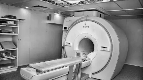
Imagine a world where nuclear medicine scans, critical for diagnosing complex conditions like cancer and heart disease, become not only more precise but also safer and more accessible to hospitals around the globe. A groundbreaking development in imaging technology, centered on the use of perovskite crystals, is poised to turn this vision into reality. Researchers have unveiled a new type of camera for single-photon emission computed tomography (SPECT), a technique that captures gamma rays from radioactive tracers to visualize organ function. This innovation, driven by collaborative efforts from leading universities, promises to overcome the persistent challenges of cost, image quality, and patient safety that have long plagued existing systems. By harnessing a material previously celebrated in solar energy, this advancement signals a potential revolution in medical diagnostics, offering sharper images with lower radiation exposure and at a fraction of the cost of current alternatives.
Breaking New Ground in Imaging Technology
Unveiling Perovskite’s Potential in SPECT
The emergence of perovskite crystals as a core component in nuclear medicine imaging marks a significant leap forward from traditional detector materials. Unlike cadmium zinc telluride (CZT), which delivers high resolution but comes with a prohibitive price tag often reaching millions of dollars, or sodium iodide, which is more affordable but yields blurrier images, perovskites offer a compelling balance of performance and cost. Specifically, cesium lead bromide (CsPbBr3) crystals have been engineered into a prototype camera that achieves an unprecedented energy resolution of 2.5% at 141 keV—a critical threshold for detecting emissions from technetium-99m, a commonly used tracer. This high sensitivity translates into the ability to distinguish tiny radioactive sources just 3.2 millimeters apart, paving the way for remarkably detailed scans that can enhance diagnostic accuracy without burdening healthcare budgets.
Beyond technical achievements, the implications of this technology extend to patient care on a profound level. With the perovskite-based camera, scans can be conducted using lower doses of radiation, minimizing exposure risks for individuals undergoing frequent imaging. Additionally, the improved resolution reduces scan times, making the process more comfortable and efficient for patients while allowing medical facilities to handle higher volumes of cases. The affordability of perovskites, due to their relatively simple growth process compared to CZT, suggests that even smaller or underfunded hospitals could soon access top-tier diagnostic tools. This democratization of advanced imaging could significantly reduce disparities in healthcare quality across different regions and economic contexts.
Engineering Excellence Behind the Innovation
Delving into the craftsmanship of this technology reveals meticulous attention to detail in the development of perovskite detectors. Researchers have focused on growing high-purity crystal boules and polishing their surfaces to near-perfect smoothness, a step that minimizes charge loss and ensures consistent performance across the detector array. These crystals are then arranged into a pixelated format, enabling uniform response and detailed image reconstruction that rivals or surpasses current standards. The integration of a custom multi-channel readout system further enhances the camera’s ability to capture precise data, setting a new benchmark for what SPECT imaging can achieve in clinical settings.
The journey from concept to prototype also highlights a remarkable cross-disciplinary approach, drawing from materials science to adapt a substance originally prominent in solar energy applications. This adaptation underscores the versatility of perovskites and their potential to address challenges beyond their initial domain. As efforts continue to refine detector design and scale up production, the focus remains on maintaining this high level of precision while ensuring the technology remains cost-effective. Such advancements suggest that the field of nuclear medicine could soon see widespread adoption of these cameras, fundamentally altering how diagnoses are performed and broadening access to cutting-edge care for diverse populations.
Looking Ahead to a New Era in Diagnostics
Enhancing Patient Outcomes with Safer Scans
Reflecting on the impact of perovskite cameras, it becomes evident that patient safety stands as a cornerstone of this technological stride. By achieving superior energy resolution and sensitivity, these detectors allow for clearer imaging with significantly reduced radiation doses, a critical factor for individuals requiring repeated scans for chronic conditions. The ability to complete scans more quickly also alleviates the physical and emotional strain on patients, transforming a once daunting procedure into a more manageable experience. This focus on minimizing harm while maximizing diagnostic precision underscores a pivotal shift in how nuclear medicine balances risk and benefit.
Moreover, the economic advantages of perovskite technology are poised to reshape healthcare delivery in profound ways. The lower production costs enable more facilities to adopt advanced imaging systems, ensuring that high-quality diagnostics are no longer a privilege reserved for well-funded institutions. This accessibility fosters a more equitable landscape, where patients in underserved areas gain access to the same level of care as those in major medical centers. Looking back, the integration of such an affordable yet powerful tool marks a turning point, addressing systemic barriers and enhancing outcomes across diverse communities.
Future Horizons for Perovskite Applications
Turning to the future, the successful deployment of perovskite cameras in SPECT imaging opens doors to broader applications and ongoing innovation. The adaptability of these crystals hints at potential uses in other medical imaging modalities or even in fields outside healthcare, such as security and industrial inspection. Continued research aimed at optimizing crystal growth and detector configurations promises to unlock even greater capabilities, potentially setting new standards for precision and efficiency in gamma-ray detection over the coming years.
As commercialization efforts gain momentum through initiatives by university spinouts, the path toward widespread adoption becomes clearer. Hospitals and clinics stand to benefit from integrating these systems, supported by scalable production methods that maintain affordability. For stakeholders in nuclear medicine, the next steps involve collaboration between researchers, manufacturers, and healthcare providers to refine implementation strategies and address any logistical challenges. This forward-looking approach ensures that the legacy of perovskite innovation will continue to evolve, ultimately delivering safer, more accurate diagnostics to patients worldwide.


