
In the critical minutes following an ischemic stroke, every second counts as brain cells begin to die, making the race against time the single most defining factor in a patient’s outcome. A landmark study by researchers Wang, D., Liang, S., and Yue, Z. illuminates a transformative advancement in emergency neurology, demonstrating how rapid neuroimaging technologies are fundamentally revolutionizing stroke care. These sophisticated diagnostic tools offer an unprecedentedly clear and immediate window into the affected brain, empowering medical teams to make life-altering decisions with greater speed and precision. This leap forward is not merely an incremental improvement; it represents a paradigm shift, moving stroke treatment from a race against a clock to a targeted, data-driven intervention that can salvage precious brain tissue and significantly improve a patient’s chances for a meaningful recovery.
The Diagnostic Power of Rapid Imaging
Seeing the Salvageable Brain
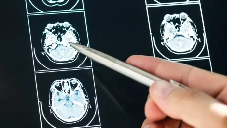
The fundamental challenge in treating an ischemic stroke, which results from an arterial blockage depriving the brain of oxygenated blood, is the rapid and unforgiving progression of cellular death. The research underscores the paramount importance of distinguishing between two critical zones within the affected brain region: the ischemic core and the surrounding penumbra. The ischemic core is the area of tissue that has already suffered severe and irreversible damage due to a profound lack of blood flow. For this central region, the window for intervention has closed. Surrounding this zone of infarction, however, is the penumbra, a region of brain tissue that is metabolically stressed and functionally impaired but remains viable. This area is the primary target of all acute stroke therapies. The cells within the penumbra are at immediate risk of dying, but if blood flow can be promptly restored, they can be saved. The study by Wang and colleagues establishes that fast imaging techniques, such as CT perfusion and advanced MRI, are indispensable for this task. These modalities provide a dynamic, functional map of cerebral blood flow, allowing clinicians to accurately delineate the boundaries of the salvageable penumbra, thereby quantifying the potential benefit of aggressive reperfusion therapies.
This ability to visualize and measure the penumbra is what transforms stroke care from a generalized emergency response into a highly personalized medical intervention. Instead of relying solely on the time since symptom onset, clinicians can use this detailed anatomical and physiological information to guide their treatment strategy. For instance, a patient with a large penumbra and a small ischemic core is an ideal candidate for interventions like intravenous thrombolysis (the administration of clot-busting drugs) or mechanical thrombectomy (the physical removal of the clot using a catheter). Conversely, a patient whose imaging reveals a large, established core with little to no surrounding penumbra may not benefit from these aggressive treatments and could be exposed to unnecessary risks, such as hemorrhage. This level of diagnostic clarity, delivered within minutes of a patient’s arrival at the hospital, empowers medical teams to make confident, evidence-based decisions. The research conclusively shows that this precision directly correlates with improved patient outcomes, minimizing long-term disability and maximizing the potential for neurological recovery by ensuring the right treatment is given to the right patient at the right time.
The Critical Role of Collateral Circulation
Beyond identifying salvageable tissue, rapid imaging provides crucial insights into a patient’s collateral circulation, a network of smaller, secondary blood vessels that can provide an alternative route for blood to reach the brain when a primary artery is blocked. The robustness of this natural bypass system is a powerful and independent predictor of clinical outcome. In patients with well-developed collateral pathways, these vessels can sustain the at-risk penumbra for a longer duration by providing a compensatory supply of blood. This effectively extends the therapeutic window, giving clinicians more time to initiate and complete reperfusion therapies. A patient with poor collateral circulation, on the other hand, will experience a much more rapid progression from ischemic penumbra to irreversible infarction, making time even more critical. Understanding the state of a patient’s collateral flow is therefore not just an academic exercise; it is a vital piece of prognostic information that shapes the entire clinical approach, from the urgency of the intervention to the prediction of the patient’s likely recovery trajectory.
The study by Wang and colleagues highlights that advanced imaging modalities are exceptionally adept at visualizing and quantifying the effectiveness of these collateral pathways. Techniques like multiphase CT angiography can map the flow of blood through these alternative routes in real-time, providing a detailed assessment of their capacity to compensate for the primary blockage. This detailed information allows for a more nuanced and personalized risk-benefit analysis for each patient. For example, a patient outside the conventional time window for treatment might still be considered a candidate for thrombectomy if imaging reveals a robust collateral network sustaining a large area of salvageable tissue. This personalized approach moves stroke care away from rigid, time-based protocols and toward a more flexible, physiology-based paradigm. By providing a comprehensive picture of both the threatened brain tissue and the body’s natural defense mechanisms, fast imaging allows clinicians to tailor their strategies, predict which patients will benefit most from intervention, and better manage expectations for long-term recovery.
From Research to Clinical Impact
Key Research Findings
The research conducted by Wang, D., and his colleagues involved a rigorous comparative analysis that pitted their newly developed fast imaging protocols against standard, more time-consuming diagnostic approaches in a cohort of acute stroke patients. The results were not merely significant; they were described as a “striking improvement” in the ability to accurately delineate salvageable brain tissue and provide a comprehensive assessment of individual collateral circulation patterns. This enhanced diagnostic precision had a direct and tangible impact on patient care. The study demonstrated that individuals who underwent the rapid imaging protocol were significantly more likely to receive timely and appropriate intervention. This correlation was not coincidental but causal; the clarity and speed of the diagnosis enabled clinical teams to proceed with confidence and urgency, which in turn translated into demonstrably improved recovery trajectories and a marked reduction in long-term disability for the patients in this group. The findings serve as powerful validation for the central thesis that speed and precision in diagnostics are inextricably linked to the preservation of brain function following a stroke.
This validation is particularly evident in its effect on one of the most critical metrics in stroke care: the “door-to-needle” time for thrombolytic therapy or the “door-to-puncture” time for mechanical thrombectomy. These metrics measure the duration from a patient’s arrival at the hospital to the initiation of treatment, and minimizing this time is a primary goal of all modern stroke centers. The study’s findings show that fast imaging protocols act as a powerful catalyst in streamlining the entire emergency response workflow. By providing definitive diagnostic and prognostic information in a fraction of the time required by older methods, these techniques eliminate delays and uncertainties in the decision-making process. This acceleration is crucial because for every minute that treatment is delayed, millions of neurons are lost. Therefore, by compressing the diagnostic timeline, rapid imaging directly contributes to better clinical outcomes, effectively operationalizing the medical axiom that “time is brain” and setting a new standard for efficient and effective acute stroke management.
Practical Advantages in Patient Care
The overarching contribution of fast stroke imaging, as synthesized in the research, lies in a powerful dual advantage that fundamentally transforms the management of acute stroke patients. Firstly, and most directly, it facilitates the timely and accurate identification of the primary therapeutic target: the salvageable penumbra. This moves the basis of treatment decisions from a reliance on often-unreliable estimations of symptom onset time to a direct visualization of the brain tissue that can still be saved. This shift to a tissue-based-window approach is a cornerstone of modern stroke care, allowing more patients to be eligible for life-changing therapies. Secondly, the technology deepens the clinical team’s understanding of the underlying individual pathophysiology by elucidating the role and robustness of the patient’s collateral circulation. This information enables the creation of a highly personalized treatment plan, one that is tailored not just to the clot itself but to the patient’s unique cerebrovascular response to the ischemic event, thereby maximizing the potential for a positive outcome.
Furthermore, the prognostic information gleaned from these advanced scans offers benefits that extend far beyond the immediate emergency room decisions. A comprehensive initial assessment of the ischemic core, penumbra, and collateral flow provides an invaluable baseline for tracking a patient’s progress and planning their long-term care. For instance, understanding the initial extent of irreversible damage can help set realistic expectations for recovery and guide the intensity and focus of rehabilitation strategies, such as physical, occupational, and speech therapy. Clinicians can better anticipate potential complications and tailor follow-up monitoring and secondary prevention efforts based on the patient’s specific vascular profile. This forward-looking perspective ensures that care is continuous and integrated, connecting the hyperacute phase of treatment with the subsequent journey of recovery. In this way, fast imaging not only saves brain tissue in the critical early hours but also provides a foundational roadmap for optimizing a patient’s quality of life for years to come.
The Future of Stroke Diagnostics
The Role of Artificial Intelligence
An overarching trend identified in the research is the progressive integration of cutting-edge technology, particularly artificial intelligence (AI), into the field of neuroimaging. The authors posit that AI and machine learning algorithms are poised to become a transformative force in the future of stroke diagnostics, amplifying the benefits of rapid imaging even further. These sophisticated computational systems can analyze complex, multi-modal imaging data with a speed and precision that surpasses human capabilities. AI algorithms can be trained to automatically identify and quantify the ischemic core and penumbra, segmenting these critical regions in a matter of seconds and providing immediate, objective data to the clinical team. This automation not only drastically accelerates the entire diagnostic workflow but also introduces a higher level of standardization and consistency, reducing the potential for inter-rater variability that can occur with manual analysis.
This synergy between advanced imaging and AI promises to unlock deeper insights from the data. Machine learning models can detect subtle patterns in cerebral blood flow, tissue health, and collateral circulation that might be imperceptible to the human eye. By integrating imaging data with other clinical information, such as patient demographics and vital signs, AI-powered software can generate more reliable predictions of patient outcomes and their likely response to specific treatments. This provides clinicians with a powerful decision-support tool, helping them to more accurately weigh the risks and benefits of interventions like thrombectomy. This forward-looking perspective situates the research within a broader technological evolution in medicine, where the partnership between human expertise and computational power is set to redefine the standards of care, making stroke treatment faster, more accurate, and more personalized than ever before.
Shaping Clinical Practice
The compelling evidence presented by Wang and colleagues did more than just validate a new technology; it provided a blueprint for the future of acute stroke management worldwide. The study’s objective demonstration of improved patient outcomes catalyzed the development and adoption of new clinical guidelines and protocols, standardizing a more effective and rapid approach to diagnosis and treatment. By solidifying the principle that timely, precise diagnostics are the absolute cornerstone of effective stroke intervention, the research helped shift the global standard of care toward a tissue-based, rather than strictly time-based, paradigm for intervention. It captured a pivotal moment in the evolution of emergency medicine, where a technological innovation directly translated into preserved neurological function and an enhanced quality of life for countless individuals affected by this life-altering condition.
This work also served as an influential call for continued research and development in the field of neuroimaging and its clinical applications. The study effectively highlighted that as synergistic technologies like machine learning and AI continue to advance, the potential to further refine diagnostic accuracy and improve patient lives will only grow. The findings solidified a new foundation upon which future innovations could be built, encouraging further exploration into automated analysis, predictive modeling, and the integration of imaging data into comprehensive digital health platforms. The legacy of this research was its clear demonstration that investing in faster, more intelligent diagnostic tools yields a profound return in the form of salvaged brain tissue, reduced long-term disability, and a more hopeful future for stroke patients and their families.