Nuclear medicine uniquely blends science and technology, reshaping patient care through the use of low-level radioactive substances. This innovative field transcends the limitations of typical radiographic techniques, such as X-rays, which only visualize structural aspects of the body. Nuclear medicine enables medical professionals to observe the functionality of organs and tissues, providing vital data for diagnosing a variety of ailments, including cardiac conditions and cancers.By facilitating the connection between diagnosis and therapy, nuclear medicine has established itself as a critical component of modern healthcare. It allows for earlier detection and more precise management of diseases, enhancing patient outcomes. This synergy of chemistry, physics, and computerized technology not only unveils the body’s internal processes but also directs clinicians toward more effective treatment paths. The diagnostic power of nuclear medicine continues to evolve, promising even greater contributions to medical science and patient care in the future.
Nature of Nuclear Medicine
Stepping into the world of nuclear medicine, one discovers that it represents an intricate blend of various scientific disciplines. It employs small amounts of radionuclides to explore the body’s labyrinth, revealing not only the structures of organs and tissues but also their dynamic functions. This ability to visualize physiological processes in real-time paves the way for a more nuanced understanding of diseases—a capability where conventional imaging techniques such as X-rays fall short. By traversing the boundaries between structure and function, nuclear medicine provides a more comprehensive picture of a patient’s medical condition, aiding in precise diagnoses and effective treatment strategies.
Radionuclide Application
In nuclear medicine, a custom radiopharmaceutical, intravenously administered, homes in on specific bodily areas for examination. As the introduced radioactive substances target certain organs or tissues, they emit radiation signatures. These are then detected by a gamma camera. The intricate process transforms emissions into detailed images, unraveling the hidden intricacies within the human body. The gamma camera’s ability to capture these emissions culminates in powerful visual data, rendering the invisible visible. This interplay is vital, paving the path for physicians to gain a profound understanding of internal conditions, and making nuclear imaging an essential instrument in medical diagnostics. The synergy of advanced imaging technology and radioactive tracers offers a non-invasive peek into the body’s secret chambers, contributing greatly to medical science’s capability to diagnose and manage various diseases.
Detection and Interpretation
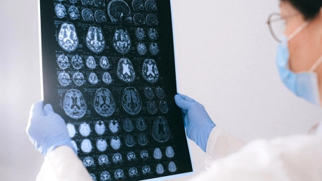
The resulting images from a nuclear scan can be likened to a celestial map, where areas of high radionuclide concentration—hot spots—shine brightly, indicating increased activity or possible pathology. Conversely, cold spots signify areas with less absorption, often suggesting a lack of function or the presence of disease. These images can vary from simple two-dimensional snapshots to complex three-dimensional reconstructions akin to CT scans, through the use of cutting-edge technologies like SPECT and PET. Such detailed views of the human body enable the detection and analysis of health-related issues with unprecedented precision, guiding medical interventions toward optimal outcomes.
Broad Applications
Nuclear medicine is at the forefront of solving myriad health issues with its specialized scans. These sophisticated tools address a wide array of medical problems through detailed imagery of specific organs. Take renal scans as an example; they’re employed to meticulously evaluate kidney functions or pinpoint blockages in blood circulation. When it comes to the thyroid gland, scans uncover vital data on its operation, aiding in the exploration of irregularities. Bone scans also prove indispensable, identifying arthritis, tumors, and tracing mysterious pain sources. These methods go beyond traditional imaging; they are instrumental in refining treatment plans, steering medical interventions, and enhancing our understanding of the intricacies of human health. Nuclear medicine, with its diverse applications, continues to illuminate the path for improved patient outcomes.
Therapeutic Uses
Beyond its diagnostic prowess, nuclear medicine also extends its virtues to the therapeutic domain, confronting maladies head-on. Conditions such as hyperthyroidism and thyroid cancer are routinely managed with its interventions. It has also become a beacon of hope for lymphoma patients and those confronting the deep-seated bone pain associated with cancer. By wielding radioactive isotopes not only as agents of visualization but also as a means of treatment, nuclear medicine showcases its dual capacity to both reveal and heal, solidifying its role in the pantheon of medical sciences.
Procedure for Performing a Nuclear Medicine Scan:
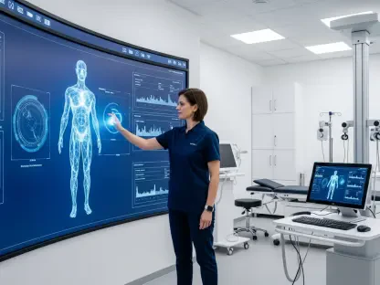
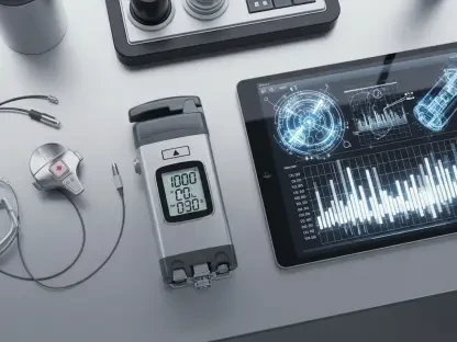
#### Tracer Administration Embarking on the path of a nuclear scan starts with the critical step of introducing a specially selected tracer. The choice of this radiopharmaceutical is strategic, tailored to the precise goals of the investigation and the particular tissues or organs of interest. Once administered, this compound commences its voyage through the patient’s body, seeking out and accumulating in regions pertinent to the scan’s focus. As the tracer circulates, it emits signals that are captured by the scanning technology, painting a picture of the body’s inner workings. This method provides a valuable window into the atomic-scale functions occurring within cells and tissues, allowing healthcare professionals to diagnose, assess, and monitor various conditions. The data gleaned from this journey is critically analyzed, for it holds clues to the mysteries of the body’s elusive ailments. This scan bridges the gap between the seen and unseen, offering a map to guide medical experts in understanding the complexity of human biology.#### Image CaptureAs a patient lies still, the silent yet powerful gamma camera operates above them, meticulously capturing the unseen dance of radioactive tracers coursing through their body. This crucial phase of data collection hinges on the patient’s immobility—every pose held is essential, contributing to the clarity and precision of the resulting images. These images are indispensable, acting as a meticulous internal map. They provide essential insights necessary for accurate diagnosis or for shaping the path of subsequent treatment. The interplay of emitted gamma rays, captured by the camera, outlines the physiological processes in detail, offering a non-invasive, yet revealing window into the body’s functioning at a molecular level. This advanced imaging technique is an invaluable tool in the medical field, bridging the gap between symptoms and diagnostics, steering medical professionals toward the correct course of action with certainty and specificity.#### Analyzing Results In the quiet orchestration of radioactive tracing, a patient’s internal workings are captured and translated into detailed images, offering a window to their internal health. Health professionals scrutinize these images—each one brimming with the possibility to shed light on an ailment, to hone in on a diagnosis, or to guide a treatment plan—all fruits of the meticulous nuclear scanning process.The time between the introduction of the tracer to the final image interpretation can vary, influenced by the specific tissue and tracer used. While some scans are swift, capturing images within moments, others require the passage of days for the full picture to emerge. Throughout this period, both patient and medical team are united in anticipation, awaiting the insights that nuclear medicine will unveil. This period is a testament to the careful balance between patient care and the complex scientific processes that underpin modern medical imaging.#### Example: Process for a Resting Radionuclide Angiogram (RNA) ScanBefore the scan, patients prepare by relinquishing metallic adjuncts and donning a hospital gown if necessary. The establishment of venous access via an IV line sets the stage for what follows. Connected to monitoring devices, the circulatory performance becomes a focal point.Tagging red blood cells with radionuclides paves the way for their subsequent deployment into the heart’s chambers. A delicate balance is struck as patients remain a silent partner in the procedure, ensuring the capture of clear images. The dance culminates with the gamma camera’s meticulous data collection.With images secured and the IV removed, the patient is free to depart, heralding the end of the nuclear sojourn. It is within the treatment centers of the Johns Hopkins network that such procedures unfold, with facilities like Howard County General Hospital, Sibley Memorial Hospital, and Suburban Hospital standing ready to guide patients on this journey through the very essence of their being.