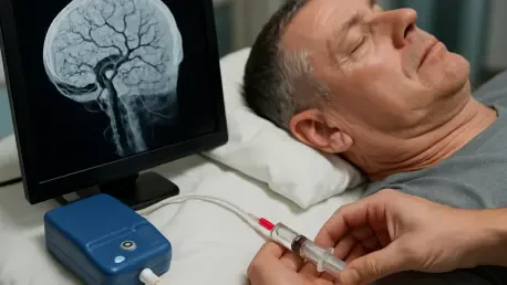
Imagine a patient who has endured persistent, debilitating headaches for years, with pain radiating from the neck to the back of the head, defying standard treatments and impacting daily life. These individuals may be suffering from cervicogenic headaches, a condition where pain originates from the cervical spine, often linked to the upper neck joints. This type of headache, though not as widely recognized as migraines, affects a significant portion of chronic headache sufferers, with prevalence estimates ranging from 0.4% to 2.5% of the general population. The complexity of the upper cervical anatomy, coupled with its proximity to critical structures like the vertebral artery and spinal cord, poses unique challenges for diagnosis and intervention. Fortunately, advancements in imaging technology, particularly CT guidance, have opened new avenues for precise and safe injections targeting the C1-C2 lateral mass joints, offering both diagnostic clarity and therapeutic relief. This article explores the anatomical intricacies, technical considerations, and procedural nuances of using CT-guided injections to address pain stemming from these joints, shedding light on a promising approach for managing a condition that has long frustrated patients and clinicians alike.
1. Understanding the Roots of Cervicogenic Headaches
Cervicogenic headaches represent a unique category of pain that stems from issues within the cervical spine, specifically tied to the upper three cervical nerve roots, and often manifesting as discomfort in the occipital or suboccipital regions. First documented in medical literature in 1983, the understanding of this condition has evolved significantly, highlighting a convergence of nerve signals from the trigeminal nerve and upper cervical nerves as a key mechanism for referred pain. The prevalence, while seemingly low at 0.4% to 2.5%, rises notably among chronic headache patients in specialized pain clinics, indicating a need for targeted awareness and intervention. Anatomically, pain sources include cervical synovial joints, muscles, intervertebral discs, and dura mater, with the C2-C3 zygapophyseal joints and C1-C2 lateral atlantoaxial joints being primary culprits. This intricate web of pain pathways underscores the importance of precise diagnostic and therapeutic strategies to address the root cause effectively.
The challenges in managing these headaches are compounded by the delicate anatomy of the upper cervical spine, where critical structures such as the vertebral artery and spinal cord lie in close proximity to potential intervention sites, creating significant concerns for practitioners. This anatomical complexity can generate apprehension among those considering invasive procedures, as even minor missteps could lead to serious complications. However, advancements in imaging, particularly CT-guided techniques, have provided a safer framework for navigating these risks. By focusing on the C1-C2 lateral mass joints, often implicated in pain generation, interventions can be tailored to confirm the pain source and provide relief, bridging the gap between conservative treatments and more invasive surgical options. This discussion sets the stage for a deeper look into the specific anatomical and technical aspects of such interventions.
2. Exploring C1-C2 Joint Anatomy
The C1-C2 articulation, also known as the atlantoaxial joint, consists of three distinct components: two synovial-lined lateral mass joints, often referred to as facet joints, and a central pivot joint formed by the dens of C2 and the arch of C1. These synovial joints can communicate anteriorly across the arch-odontoid articulation, which influences how contrast spreads during diagnostic or therapeutic injections. The combined volume capacity of these joints typically ranges from 3.5 to 4 mL when they communicate, dropping to about half that amount when isolated. This structural detail is critical for practitioners to understand when planning the volume of injectate to avoid overfilling or extravasation, which could lead to unintended complications or reduced efficacy of the procedure.
Pathologically, the C1-C2 lateral mass joints are susceptible to conditions affecting any synovial-lined joint, though serious issues like infections, inflammatory diseases such as rheumatoid arthritis or gout, and malignancies are rare. More commonly, pain in these joints arises from degenerative changes or post-traumatic arthrosis, conditions that wear down the joint surfaces over time or following injury. These degenerative processes can alter the joint’s capacity and response to injections, necessitating careful preprocedural imaging to assess the extent of damage. Understanding these anatomical and pathological nuances is foundational for safely targeting the C1-C2 joints with CT-guided injections, ensuring that interventions are both precise and tailored to the patient’s specific condition.
3. Innervation and Pathological Insights of C1-C2 Joints
The innervation of the C1-C2 facet joints is complex, with the dorsolateral aspects receiving input from 5 to 9 articular branches stemming from various segments of the C2 nerve, including the dorsal root ganglion, ventral spinal nerve, and ventral ramus. A prevertebral nerve plexus also contributes to this network, adding to the intricacy of pain transmission in this region. The C2 dorsal root ganglion, positioned at the neural foramen near the dorsal margin of the lateral mass articulations, is particularly sensitive and can be inadvertently stimulated during posterior approaches, potentially causing significant discomfort. This anatomical detail emphasizes the need for meticulous needle placement to avoid unwanted nerve irritation during injections.
Pathologically, stimulation of the C1-C2 facet joints through contrast injection often results in suboccipital or occipital pain, while diagnostic injections with anesthetics have proven effective in alleviating such symptoms, confirming the joint as a pain source in many cases. Studies suggest that lateral atlantoaxial joint pathology contributes to approximately 16% of occipital headaches, with osteoarthritis and trauma being the predominant causes. Imaging reveals characteristic degenerative signs like joint space narrowing, subchondral sclerosis, and cysts on CT, alongside bone marrow edema on MRI. The prevalence of atlantoaxial arthritis increases with age, affecting up to 18.2% of individuals in their later decades, highlighting the importance of age-specific considerations in treatment planning for cervicogenic headaches.
4. Pain Patterns and Therapeutic Interventions for C1-C2 Joints
Pain associated with cervicogenic headaches originating from the C1-C2 joints is often variable and subjective, frequently radiating to the frontal, temporal, or occipital regions, and may present unilaterally or bilaterally. This variability complicates clinical diagnosis, as physical examination findings struggle to precisely localize the pain source due to overlapping pathologies and intricate innervation patterns in the upper cervical spine. Standardized diagnostic criteria from the International Headache Society help by integrating clinical, laboratory, and imaging evidence to confirm a cervical origin, often requiring controlled diagnostic blocks to pinpoint the exact pain generator. This diagnostic challenge necessitates advanced imaging and intervention techniques to achieve accurate results.
Therapeutic options for managing C1-C2 joint pain span a wide spectrum, each with varying degrees of invasiveness and efficacy. Conservative measures, such as physical therapy and anti-inflammatory medications, offer minimal to moderate relief for many patients. On the other end, surgical interventions like C1-C2 arthrodesis provide long-term pain reduction—sometimes exceeding two years—but limit head rotation and carry significant risks. Injections directly into the C1-C2 facet joints have shown promising results, with 81% of patients experiencing at least a 50% pain reduction lasting up to three months. Alternative approaches, including neurectomy of occipital nerves or ligation of adjacent arteries, report relief in 86% of cases, though these lack specific focus on C1-C2 joint pathology in their assessments, underscoring the need for targeted interventions like CT-guided injections.
5. Indications for Targeting C1-C2 Joints with Injections
The primary purpose of C1-C2 lateral mass articulation injections is diagnostic, aimed at determining whether these joints are the source of a patient’s headaches after other potential causes, such as general facet arthrosis or migraines, have been ruled out. Imaging plays a pivotal role in this process, with MRI being particularly useful for detecting signs of edema or joint effusions that suggest active pathology. If these indicators are absent, it is less likely that the C1-C2 joint is the pain generator, guiding clinicians away from unnecessary procedures. This step of exclusion and confirmation is crucial for ensuring that interventions are both justified and likely to yield meaningful results for the patient.
Patients often present with symptoms mimicking occipital neuralgia, typically on the same side as the affected joint or where impingement of the C2 dorsal root ganglion occurs, adding to the diagnostic complexity. Due to the inherent risks of accessing this anatomically sensitive area, many practitioners opt to include steroids in diagnostic injections to provide a longer-lasting therapeutic effect. This dual-purpose approach, however, makes it challenging to discern whether immediate relief stems from the local anesthetic or the delayed action of the steroid, which can take up to 14 days to fully manifest. Pain levels are meticulously documented before and after the procedure using numerical scales and diaries, with a 50% reduction in pain considered a positive response, reflecting the unique anatomical considerations of this joint compared to other cervical facets.
6. Comparing Fluoroscopy and CT Guidance for Injections
Fluoroscopy has long been a standard method for guiding C1-C2 lateral mass joint injections, with well-documented techniques relied upon by experienced interventionalists for their accessibility and real-time imaging capabilities. This approach allows for dynamic visualization of needle placement under X-ray, providing immediate feedback during the procedure. While effective in many settings, fluoroscopy can sometimes lack the depth perception and detailed soft tissue contrast needed to navigate the complex anatomy surrounding the upper cervical spine, potentially increasing the risk of misplacement or injury to nearby structures like the vertebral artery.
In contrast, CT guidance offers distinct advantages by enabling a stepwise assessment of needle depth and precise identification of soft tissue structures in relation to the needle tip in real-time, which is crucial for ensuring accuracy during delicate procedures. This enhanced precision is particularly beneficial when targeting the narrow safe corridor of the C1-C2 joint, reducing the likelihood of complications. However, limitations exist, such as beam hardening artifacts caused by dental amalgam or implanted spinal hardware, which can obscure imaging. These challenges are often mitigated through metal artifact reduction techniques, such as adjusting kV and mAs settings, to improve image quality. The choice between these modalities often depends on specific patient anatomy, equipment availability, and practitioner expertise, with CT guidance emerging as a powerful tool for enhancing procedural safety.
7. Essential Preprocedural Evaluations
Before proceeding with a C1-C2 lateral mass articulation injection, identifying these joints as the probable source of pain is paramount, achieved through comprehensive clinical examinations, detailed imaging studies, and the systematic exclusion of alternative diagnoses. This initial step ensures that the intervention is appropriately targeted, avoiding unnecessary risks in cases where other conditions might be responsible for the symptoms. Determining whether the injection serves a diagnostic purpose—to confirm the pain source—or a therapeutic one—to provide relief—is also critical in shaping the procedural approach and patient expectations.
Several additional factors must be addressed during preprocedural planning to maximize safety and efficacy, ensuring that all potential risks are mitigated. Absolute contraindications, as outlined by the Spine Intervention Society for fluoroscopically guided injections, apply equally to CT-guided procedures and must be strictly adhered to. Informed consent discussions cover the risks, benefits, alternative treatments, and expected recovery process, ensuring patients are fully aware of the procedure’s implications. A thorough review of the patient’s medications and comorbidities is necessary, particularly regarding anticoagulant use, which could heighten bleeding risks, and glycemic control if steroids are planned, due to potential systemic absorption effects. This meticulous preparation forms the foundation for a safe and effective intervention, tailored to the individual’s medical profile.
8. Selecting the Appropriate Injectate
For purely diagnostic injections targeting the C1-C2 joints, options include 1% lidocaine, which provides an immediate but short-acting effect, or 0.25% bupivacaine, offering analgesia for approximately five hours. The former is often preferred in outpatient settings due to its rapid onset and brief duration, allowing for quick assessment of pain response without prolonged monitoring. There is no definitive evidence in the literature favoring one over the other, so the choice often hinges on procedural context and patient needs, ensuring that the diagnostic window is both practical and informative for confirming the pain source.
When therapeutic relief is also a goal, a combination of 0.5 mL of 1% lidocaine with 1 mL of dexamethasone at 10 mg/mL is commonly used as the initial injectate, leveraging the benefits of immediate anesthesia and longer-term anti-inflammatory action. Non-particulate steroids like dexamethasone are favored in the cervical region to minimize the catastrophic risk of intravascular injection leading to brain embolization. If a patient has previously responded well to this combination but experiences recurrent pain, the risks and benefits of using a particulate steroid, such as triamcinolone acetate, may be discussed to potentially achieve more sustained relief. This tailored approach to injectate selection balances diagnostic precision with therapeutic outcomes, prioritizing patient safety in a high-risk anatomical area.
9. Navigating Anatomical Risks and Proximities
The C1-C2 lateral mass joint is surrounded by critical structures that pose significant risks during injections, requiring a thorough understanding of anatomical proximities to ensure a safe trajectory. Medially, the thecal sac and its contents are at risk, where a deviated needle path could result in intrathecal injection, spinal cord or nerve root injury, or cerebrospinal fluid leak. Laterally, the vertebral artery presents a hazard for intravascular injection or catastrophic hemorrhage, particularly in patients on anticoagulation therapy, with its position varying significantly among individuals. Prior imaging must be reviewed to map out these variations and plan the safest approach.
Ventrally, advancing the needle too far risks encountering the internal carotid artery or even extending into the oropharynx, while dorsally, the C2 dorsal root ganglion and ventral ramus are highly sensitive to mechanical stimulation, potentially causing intense pain if contacted. Additionally, a venous plexus in the dorsal region could lead to bleeding complications in patients with coagulopathy. To mitigate these risks, needle advancement should be performed in short, intermittent steps, with small doses of 1% lidocaine administered near the posterior joint margin to reduce discomfort from nerve contact. This cautious approach, combined with real-time CT imaging, helps avoid critical structures and enhances procedural safety in this anatomically confined space.
10. Positioning Patients for CT-Guided Procedures
Proper patient positioning is a cornerstone of successful CT-guided C1-C2 injections, ensuring both safety and accuracy during the procedure. Patients are typically placed prone on a specialized table support designed to maximize comfort and maintain a neutral neck position, minimizing lateral rotation or flexion that could alter anatomical alignment. This setup also helps limit patient movement, which is critical for maintaining consistency between scout imaging and needle placement. Fiducial markers, radiopaque bars oriented craniocaudally, are placed on the posterior upper neck to guide initial imaging and trajectory planning, with strict instructions for the patient to remain still throughout.
Additional considerations in positioning include managing potential interferences, such as hair that might disrupt fiducial marker placement, often addressed by using a bonnet to isolate the procedural field. Scout imaging, covering the area from the odontoid to the inferior endplate of C2, is conducted in both anteroposterior and lateral views to establish a clear field of view for planning. Local anesthesia with 1% lidocaine is standard, though conscious sedation may be offered for particularly anxious patients, provided trained staff and continuous hemodynamic monitoring are available. General anesthesia is avoided to allow for immediate post-injection pain response evaluation, ensuring that diagnostic feedback is not obscured. These steps collectively create a stable and controlled environment for precise intervention.
11. Acquiring CT Images for Precision
CT image acquisition for C1-C2 injections begins with a preliminary scan using specific settings to optimize visualization: a slice thickness of 1.25 mm, 120 kVp, a tube current of 100 mA, a focal spot of 1.2, a revolution time of 600 ms, and an exposure of 3 mSv. These parameters ensure detailed imaging of the upper cervical anatomy, which is crucial for identifying the safest posterior trajectory to the target joint. Axial images are meticulously reviewed to draw a straight line from the skin surface to the posterior atlantoaxial joint opening, targeting the junction of the lateral third and medial two-thirds, while measuring the depth to select an appropriately sized needle for the procedure.
Documentation during this phase is vital, with the specific slice number recorded and a still image saved, marking the intended needle puncture site relative to the fiducial marker’s position for medial-lateral orientation. A procedural time-out is conducted to confirm all details before starting. Sequential imaging during the injection often uses 2.5 mm slices to accommodate the small size of the lateral atlantoaxial joint and navigate around potential osteophytic growths common in degenerative conditions. This real-time, high-resolution approach allows for continuous adjustment of the needle path, avoiding critical structures and ensuring that the intervention remains within the narrow safe corridor of the C1-C2 joint, enhancing both safety and accuracy.
12. Managing Contrast Use and Reactions
The use of contrast in CT-guided C1-C2 injections carries risks similar to those outlined in standard radiology guidelines, including physiologic, chemotactic, and allergic reactions, though the likelihood remains low. Patients must be questioned about prior contrast exposure and any adverse reactions to anticipate potential issues. The volume of contrast used is minimal, typically 1 to 2 mL of a nonionic monomeric agent like Omnipaque 180, starting with 0.5 to 1 mL increments to opacify the ipsilateral joint. In approximately 18% of cases, contrast may spread centrally to the pivot joint or contralateral lateral atlantoaxial joint, with dorsal and ventral recesses becoming visible, aiding in confirming correct needle placement.
For patients with documented contrast allergies, the severity of past reactions must be assessed, adhering to established protocols that may include premedication or procedural avoidance to ensure safety. Alternatives such as air or gadolinium are considered, though each carries distinct risks—air injection risks posterior circulation stroke if intravascular, while gadolinium may cause nephrogenic systemic fibrosis or other systemic effects, though it is less allergenic. Techniques like reducing kVp to 70 optimize gadolinium opacification, and CT-fluoroscopy or step-and-shoot imaging monitors for extravasation. Comparing pre- and post-contrast images at the same level ensures accurate assessment of spread, maintaining procedural safety in this delicate anatomical region.
13. Tools and Technical Considerations for the Procedure
Standard kits for CT-guided C1-C2 injections are equipped with a range of syringes, needles, and sterile preparatory materials, including non-latex gloves to prevent allergic reactions during the procedure. Spinal needles, either 3.5-inch 25-gauge or 22-gauge, are typically used, with the latter preferred for better tactile feedback and ease of redirection. If the planned trajectory exceeds 10 cm, a 5-inch needle is selected, though this is rare in cervical interventions. For diagnostic aspirations involving more viscous joint fluid, a 20-gauge needle may be necessary. The beveled tips of spinal needles allow for precise manipulation, making them suitable for both injection and aspiration tasks in this confined space.
Technical nuances during the procedure include strategies for navigating anatomical obstacles like osteophytic ridging at the posterior joint capsule, often managed by applying gentle pressure with alternating clockwise and counterclockwise rotation to access the joint. Needle advancement under intermittent CT guidance is performed in small increments of 0.5 to 1 cm, with imaging between steps to confirm trajectory and avoid critical structures. This methodical approach ensures precision, as the shadow of the needle tip often serves as a reliable guide. Such careful handling of tools and techniques minimizes the risk of complications, allowing for effective targeting of the C1-C2 lateral mass articulation while maintaining patient safety throughout the intervention.
14. Step-by-Step Technique for CT-Guided Injections
Step 1: Marking the Entry Site
The procedure begins by activating the CT scanner’s laser to mark the needle puncture site, counting to the relevant fiducial marker and drawing two lines—one craniocaudal along the closest marker and another at 90 degrees from medial to lateral based on the laser. This ensures alignment with the preplanned trajectory, which is critical for maintaining accuracy in this narrow anatomical corridor.
Step 2: Sterilization
The skin over the marked entry site is sterilized using two chlorhexidine sticks, and it is allowed to dry completely to ensure proper preparation for the procedure.
