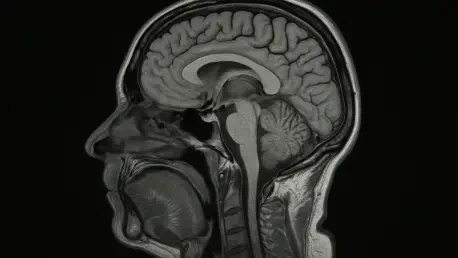
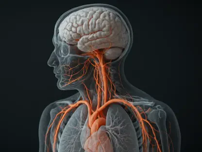
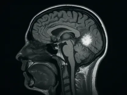
In a world where aging is usually counted in birthdays yet felt in daily clarity, strength, and recall, a new imaging thread pulled at midlife health suggested the brain’s clock might also be written in muscle and deep belly fat, not just in gray matter volume or cognitive tests alone. A research team analyzing healthy adults used whole-body and brain MRI to look beyond the bathroom scale, separating visceral adipose tissue from the softer satchel of subcutaneous fat and pairing those body maps with an AI estimate of brain age. The central question was both intuitive and high-stakes: do stronger bodies carry more resilient brains, and does hidden abdominal fat quietly advance cerebral wear? Early answers pointed to a measurable link that favors muscle and flags visceral fat as a risk, reinforcing a shift toward composition-aware prevention rather than weight alone.
What The Scans Revealed
The study evaluated 1,164 adults with a mean age near 55 using whole-body coronal T1-weighted MRI to quantify total normalized muscle volume and distinguish visceral from subcutaneous fat, then paired those metrics with a separate brain T1 volumetric scan. A fully convolutional network produced a brain age estimate, enabling a brain age gap measure defined as brain age minus chronological age. Higher muscle volume correlated with both younger ages in the cohort and younger-appearing brains, reflected by smaller or more favorable brain age gaps. In contrast, greater visceral fat relative to muscle tracked with older ages and older-appearing brains. The convergence of 3D segmentation and AI modeling turned disparate signals into a coherent marker of body–brain health, raising precise questions about which levers—muscle gain or visceral fat loss—matter most for the cortex.
What The Scans Revealed
The findings aligned with a growing consensus: body composition, not just body weight, shapes brain aging trajectories. Visceral adiposity has been linked to inflammatory signaling and metabolic stress tied to neurodegeneration risk, while muscle may confer vascular and endocrine benefits that support brain integrity. Yet the data were observational and presented as ongoing research, so causation remained unproven and generalizability beyond midlife, healthy adults must be tested. The promise lay in scalability—automated MRI segmentation and AI-derived brain age provide repeatable biomarkers that could guide individualized prevention. That roadmap pointed toward interventions that prioritize visceral fat reduction and muscle preservation, including lifestyle programs and therapies such as GLP-1–based regimens, monitored by longitudinal MRI to verify impact on brain aging profiles. Such a strategy emphasized actionable, measurable change and laid groundwork for targeted trials that could resolve causality and dosage.


