
In courtrooms and at border crossings worldwide, determining an individual’s true chronological age can be a pivotal factor that alters the course of a life, yet the methods used have long been fraught with significant ethical and medical concerns. A pioneering study from Ottow and colleagues introduces a groundbreaking technique using low-field magnetic resonance imaging (MRI) that promises a non-invasive, radiation-free solution. This innovative approach, which focuses on the developmental stages of the wrist bone, known as the distal radius, directly confronts the challenges of traditional forensic practices. By demonstrating a safe, accessible, and accurate alternative, this research stands poised to revolutionize how age is determined in critical legal scenarios, from asylum claims and immigration proceedings to criminal investigations involving minors, offering a future where scientific precision and human dignity are no longer at odds.
The Search for a Safer Method
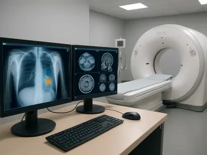
The critical need for accurate age determination permeates various legal frameworks, often becoming the deciding factor in cases of asylum, immigration disputes, and the prosecution of young offenders. For decades, forensic specialists have leaned on established techniques such as X-ray imaging to assess skeletal maturity or histological analysis of teeth and bones. However, these conventional methods are burdened by substantial drawbacks that raise serious questions. X-ray-based assessments, for example, subject individuals—frequently children and adolescents—to ionizing radiation. This exposure carries inherent health risks, prompting ethical objections that severely limit its application, particularly when repeated examinations are necessary. Other common methods can be highly invasive, requiring the extraction of tissue samples, which introduces a separate layer of legal and ethical constraints, making the process both physically and procedurally intrusive for the subject. The persistent reliance on these flawed techniques has created an urgent, long-standing demand for a safer, more humane, and scientifically robust alternative in forensic science.
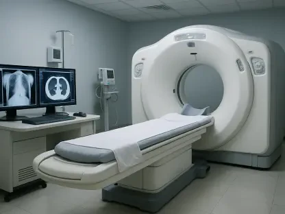
To address these profound challenges, the research team championed a novel approach centered on low-field MRI technology. Operating at a magnetic strength of 0.31 Tesla, this system is considerably less powerful than the high-field scanners typically found in clinical settings, which operate at 1.5 to 3 Tesla. While a lower magnetic field inherently presents a technical hurdle—specifically, a reduced signal-to-noise ratio that could compromise image clarity—it simultaneously offers a suite of compelling practical advantages. These benefits create a strong consensus on the technology’s vast potential, including significantly reduced acquisition and operational costs, a smaller physical footprint, less complex infrastructural demands like cooling systems, and enhanced portability. The researchers adeptly navigated the issue of image quality by developing meticulously optimized imaging protocols. Their work successfully demonstrated that even with a lower field strength, it is possible to capture images with sufficient resolution and contrast to perform detailed and reliable forensic analysis, striking an ideal balance between technological accessibility and diagnostic precision.
From Research to Real-World Impact
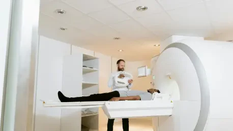
The methodology of the study is strategically anchored in the selection of the distal radius—the part of the forearm bone closest to the wrist—as its primary anatomical focus. This choice was highly deliberate, as the distal radius undergoes a well-documented and predictable series of physiological changes throughout adolescence and into early adulthood, making it a reliable barometer of skeletal maturation. Key developmental markers in this region, such as the state of the growth plate, the progression of ossification centers, and evolving cartilage patterns, provide consistent and quantifiable indicators of an individual’s age. By concentrating on these dependable markers, the research team was able to capture a comprehensive set of detailed images from a diverse cohort of living subjects spanning a broad age spectrum. The subsequent analysis involved a rigorous assessment of key morphological features that correlate strongly with age, particularly the degree of growth plate fusion and the advancement of bone formation, creating a robust framework for estimation.
The principal finding emerging from this comprehensive research is the definitive validation of the method’s viability and remarkable reliability. The study uncovered a strong and statistically significant concordance between the age-related parameters observed in the low-field MRI scans and established age benchmarks from existing medical literature. This powerful correlation confirms that 0.31 Tesla MRI of the distal radius is an effective and accurate tool for forensic age estimation, positioning it as a formidable alternative to conventional radiation-based methods. This discovery represents a major technical breakthrough, proving that the numerous advantages of low-field systems—such as affordability and portability—do not have to be traded for diagnostic accuracy. The implications of this research are profoundly transformative, heralding a potential paradigm shift toward more ethical, accessible, and equitable forensic practices on a global scale. This new technique is especially well-suited for use on minors and vulnerable populations, as it completely eliminates the health risks associated with radiation.
Navigating Challenges and Future Innovations
While the results are undeniably promising, the authors maintain a commendably objective viewpoint by openly acknowledging the study’s inherent limitations. The primary constraint identified is the lower image resolution of low-field MRI when compared directly to its high-field counterparts. This difference, while managed effectively through optimized protocols, means that finer anatomical details may be less distinct. Consequently, the researchers strongly emphasize the critical need for comprehensive validation studies involving larger and more demographically diverse populations. Such extensive research is crucial to fine-tune the diagnostic criteria and rigorously account for the natural variations in skeletal development that are influenced by a range of factors, including ethnicity, nutritional status, and overall health. Addressing these variables is essential to prevent the introduction of potential biases and to ensure the method’s broad, equitable, and consistent applicability across different global populations, thereby strengthening its legal and scientific standing.
The work of Ottow and his colleagues presented convincing evidence that 0.31 Tesla low-field MRI of the distal radius was a viable, ethical, and accurate method for forensic age estimation. This development marked a significant milestone, promising to enhance the safety, accessibility, and reliability of forensic investigations worldwide. The research also highlighted a forward-looking perspective, proposing the integration of artificial intelligence and advanced image processing to further augment the technique’s accuracy and efficiency. It was suggested that machine learning models, trained on extensive datasets of annotated MRI scans, could automate the identification of ossification stages and other critical developmental markers. This automation would not only expedite the analysis process but also significantly reduce the subjective variability that can exist between different forensic experts, paving the way for more standardized and objective outcomes. This vision underscored a growing trend of interdisciplinary collaboration, merging radiology, computer science, and forensic medicine to tackle complex medico-legal challenges and advance a future of forensic science that prioritizes scientific precision while upholding human dignity and social justice.