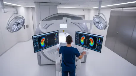

The diagnostic journey for patients with suspected renal cell carcinoma often begins with a complex trade-off between the necessity of surgical intervention and the risk of over-treating benign or indolent lesions. Traditional imaging modalities like computed tomography and standard magnetic resonance imaging frequently struggle to differentiate between low-grade tumors and aggressive malignancies without the use of invasive biopsies or contrast agents that may pose risks to patients with compromised kidney function. Magnetic Resonance Fingerprinting (MRF) has emerged as a groundbreaking quantitative imaging technique that challenges this status quo by utilizing pseudorandom pulse sequences to create unique signal signatures for different tissue types. By matching these signals against a predefined dictionary of physical properties, MRF allows for the rapid and simultaneous acquisition of multiple tissue parameters such as T1 and T2 relaxation times within a single 15-second breath-hold. This approach not only streamlines the clinical workflow but also provides a more objective, data-driven foundation for assessing the biological behavior of renal masses before a scalpel ever touches the patient.
Quantitative Metrics and Study Methodology: A New Standard
Recent prospective research has pivoted toward evaluating how effectively MRF-derived metrics can categorize the aggressiveness of renal neoplasms in adult patients slated for surgical resection. By leveraging high-field 3 T MRI scanners and noncontrast protocols, investigators have successfully generated comprehensive quantitative maps that highlight the underlying physiological differences between indolent and high-grade tumors. This specific proof-of-concept study compared MRF performance against established methods like diffusion-weighted imaging and arterial spin labeling, which measure the apparent diffusion coefficient and renal blood flow, respectively. Unlike these traditional techniques, which often require separate, time-consuming acquisitions and complex post-processing alignment, MRF offers inherent coregistration of data points. This synchronization ensures that the T1 and T2 relaxation times are measured from the exact same spatial location simultaneously, significantly reducing the errors associated with patient movement or slight changes in positioning during the imaging session.
The integration of MRF into the diagnostic pipeline represents a significant shift from qualitative visual assessments toward precise numerical characterization of tumor biology. While conventional MRI relies on the radiologist’s interpretation of signal intensity variations, MRF provides absolute values that reflect cellular density, water content, and the micro-environmental architecture of the tissue. In the analyzed patient cohort, T2 relaxation time stood out as a particularly potent indicator of malignancy grade, showing a clear statistical divergence between indolent tumors, which averaged 86 ms, and aggressive ones at 61 ms. This sensitivity to the subtle shifts in tissue composition suggests that MRF can capture the increased complexity and overcrowding of cells typical of high-grade renal cell carcinoma. Furthermore, because the technique is noninvasive and does not require exogenous contrast, it serves as a safer alternative for patients who may be at risk for nephrogenic systemic fibrosis or other complications related to gadolinium-based agents used in standard scans.
Clinical Implications and Future Directions: Refining Patient Management
Analytical models combining MRF parameters have demonstrated a remarkable ability to predict tumor behavior, often outperforming multi-modal approaches that incorporate diffusion or perfusion data. For instance, a multivariable model utilizing both T1 and T2 values achieved a robust area under the receiver operating characteristic curve of 0.89, showcasing a sensitivity of 86 percent and a specificity of 93 percent for identifying aggressive malignancies. Interestingly, the addition of renal blood flow or diffusion metrics did not provide a statistically significant boost to the diagnostic accuracy of the MRF-based model. This finding suggests that the inherent physical properties captured by MRF may be sufficient on their own to characterize the pathology of renal masses. By reducing the need for multiple specialized sequences, clinics can potentially decrease the time patients spend in the scanner while increasing the reliability of the results. This efficiency is crucial for high-volume oncology centers where rapid, accurate staging is the cornerstone of effective treatment planning.
As medical institutions move toward more personalized treatment strategies, the role of quantitative imaging in distinguishing tumor phenotypes will likely expand from 2026 to 2030. Clinicians should consider integrating MRF into standard protocols for renal mass characterization, especially when traditional imaging remains ambiguous or when biopsy is contraindicated. Future efforts must focus on validating these findings through larger, multi-center trials to account for technical variability across different scanner manufacturers and diverse patient populations. Enhancing the automated segmentation of regions of interest would also reduce inter-operator variability, ensuring that these quantitative metrics are reproducible in real-world clinical settings. The transition from purely anatomical imaging to physiological profiling through MRF provided a viable pathway for refining surgical candidacy and monitoring patients on active surveillance. Ultimately, the successful deployment of these noninvasive tools allowed for a more nuanced understanding of tumor aggressiveness, leading to improved outcomes by ensuring that aggressive interventions were reserved for the most high-risk cases.