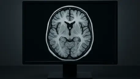
Brain MRI stands as a cornerstone in diagnosing neurological disorders, ranging from acute strokes to insidious tumors, yet it grapples with a fundamental challenge that has persisted for decades: striking a perfect balance among acquisition time, image resolution, and signal-to-noise ratio (SNR), often dubbed the “magic triangle.” In critical medical scenarios, lengthy scan durations can delay life-saving decisions, while attempts to accelerate the process frequently compromise the clarity or detail necessary for accurate assessments. Deep learning (DL), a cutting-edge subset of artificial intelligence, emerges as a potential game-changer in this arena, promising to reconstruct high-quality images from limited data through sophisticated neural networks. This technology could redefine the boundaries of MRI by harmonizing the competing elements of the magic triangle, potentially slashing scan times without sacrificing diagnostic precision. The implications are profound, particularly in emergency settings where every second counts, envisioning a future where brain imaging is both rapid and reliable. This exploration delves into recent advancements and research to evaluate whether DL can truly transform brain MRI, addressing longstanding limitations and paving the way for enhanced clinical outcomes. Can this innovative approach finally achieve the elusive synergy of speed, sharpness, and clarity that has long been sought in medical imaging?
Unpacking the Magic Triangle Challenge in Brain MRI
Brain MRI remains an indispensable tool for identifying a spectrum of neurological issues, such as ischemic changes or subtle lesions, but its prolonged acquisition times often hinder its effectiveness, especially in urgent situations. In cases like acute stroke, where rapid diagnosis can dictate the success of interventions, waiting upwards of 13 minutes for a full scan can be detrimental to patient outcomes. This delay not only affects clinical decision-making but also poses challenges for individuals who struggle with remaining still, increasing the risk of motion artifacts that obscure critical details. The core issue lies in the inherent trade-offs of the magic triangle, where optimizing one aspect—such as reducing scan time—typically degrades another, like resolution or SNR. Traditional acceleration techniques, including parallel imaging and compressed sensing, have been deployed to mitigate these delays, yet they often result in noisier or less detailed images, undermining diagnostic confidence. This persistent dilemma underscores the urgent need for a solution that can reconcile these competing demands without compromise, setting the stage for innovative approaches to redefine the capabilities of brain MRI in modern healthcare settings.
The concept of the magic triangle encapsulates the ideal scenario in brain MRI: achieving swift scans, high-resolution visuals, and minimal noise simultaneously. Historically, conventional methods have struggled to attain this balance, as enhancing one parameter invariably weakens another, creating a frustrating cycle of trade-offs. For instance, cutting acquisition time often means acquiring less data, which can blur fine anatomical structures or amplify background noise, making it harder for radiologists to discern subtle abnormalities. This challenge is particularly pronounced in emergency departments and with vulnerable patient populations, such as children or those with anxiety, where shorter scan durations are crucial for both comfort and accuracy. Enter deep learning, which offers a novel pathway by leveraging algorithms trained on extensive imaging datasets to reconstruct detailed images from incomplete or undersampled data. This technology holds the potential to break the traditional constraints of MRI, promising a future where the magic triangle is no longer an unattainable goal but a standard of care, transforming how neurological diagnostics are performed across diverse clinical environments.
How Deep Learning Could Transform MRI Reconstruction
Deep learning, a branch of artificial intelligence that employs neural networks to emulate human cognitive processes, presents a groundbreaking method for enhancing brain MRI by reconstructing high-quality images even when data collection is limited. These algorithms are trained on vast repositories of imaging data, learning intricate patterns that allow them to fill in gaps from undersampled scans, effectively producing visuals with clarity comparable to those from longer, traditional acquisitions. Unlike older acceleration techniques that often degrade image quality, DL focuses on post-processing enhancements, refining details and reducing noise after the scan is complete. This capability could fundamentally alter the MRI workflow, enabling radiologists to obtain diagnostic-grade images in a fraction of the usual time. The significance of this advancement lies in its potential to address the core limitations of brain imaging, offering a tool that prioritizes both efficiency and precision in a field where such a balance has been historically elusive.
Recent investigations, including pivotal research conducted at Mansoura University, have put DL tools like Subtle MR software to the test in real-world brain MRI applications on a 1.5 Tesla scanner. The objective was to drastically reduce scan times while preserving or even improving key image metrics like SNR and resolution. Initial findings suggest that DL can indeed refine images post-acquisition, smoothing out noise and enhancing anatomical detail without requiring extended data collection periods. This approach contrasts sharply with conventional methods, which often necessitate a direct trade-off between speed and quality. The promise of such technology extends beyond mere technical improvement; it hints at a broader shift in radiology practices, where shorter scan durations could become the norm, easing patient burden and streamlining hospital operations. However, the question remains whether these early successes can scale across diverse settings and consistently meet the rigorous demands of clinical diagnostics.
Insights from Cutting-Edge Research on Deep Learning
A comprehensive retrospective study carried out between 2022 and 2024 at Mansoura University provides compelling evidence of deep learning’s impact on brain MRI, utilizing a 1.5 Tesla scanner to compare traditional and DL-enhanced imaging protocols. The study involved 50 patients, evenly divided into two groups: one subjected to standard MRI techniques with scan times averaging 13 minutes, and the other undergoing a faster protocol of approximately 6 minutes, followed by DL reconstruction to boost image quality. The disparity in acquisition time alone highlights the potential for significant efficiency gains, particularly in high-pressure medical environments where rapid results are paramount. Beyond mere speed, the research meticulously evaluated both quantitative and qualitative outcomes to assess whether DL could uphold the standards necessary for accurate neurological assessments, offering a robust foundation for understanding its practical utility in clinical settings.
Delving into the results, the study revealed that DL-reconstructed images exhibited a remarkable improvement in SNR, particularly in fluid-attenuated inversion recovery (FLAIR) sequences, which are vital for detecting subtle brain abnormalities like white matter lesions. This enhancement in SNR translates to clearer images with reduced background noise, despite the drastically shortened scan times, suggesting that DL can maintain or even elevate diagnostic precision under accelerated conditions. Additionally, qualitative evaluations by seasoned neuroradiologists underscored the superiority of DL images, with higher ratings for anatomic detail, overall quality, diagnostic confidence, and minimized artifacts. The consistency of these assessments was further validated by strong inter-observer agreement, indicating that DL enhancements do not introduce subjective discrepancies that could compromise interpretation. These findings collectively paint a promising picture of DL as a tool capable of redefining the standards of brain MRI efficiency and reliability.
Clinical Advantages and Wider Implications of Deep Learning
The integration of deep learning into brain MRI protocols offers transformative clinical advantages, chief among them the ability to halve scan times from 13 to 6 minutes, as demonstrated in recent studies. This reduction is particularly impactful in emergency contexts, such as acute stroke management, where swift imaging can accelerate critical decisions like initiating thrombolytic therapy within narrow therapeutic windows. Faster scans also mitigate delays in busy hospital settings, ensuring that more patients can be assessed promptly, which is essential for optimizing outcomes in time-sensitive conditions. Moreover, the efficiency gained from quicker imaging protocols can alleviate bottlenecks in radiology departments, allowing healthcare providers to address patient needs with greater agility. This technical leap forward holds the promise of reshaping emergency care by prioritizing speed without undermining the quality of diagnostic insights.
Beyond immediate clinical benefits, deep learning in MRI significantly enhances patient experiences by shortening scan durations, which is a boon for those who find prolonged imaging sessions uncomfortable or distressing. Individuals with anxiety, claustrophobia, or physical limitations often struggle to remain still during traditional scans, increasing the likelihood of motion artifacts that can obscure vital details. By cutting scan times, DL reduces these risks, leading to clearer images and more reliable diagnoses while improving overall comfort. Additionally, the technology’s ability to boost scanner throughput translates to shorter wait times for patients and reduced operational costs for facilities, fostering a more streamlined healthcare system. This dual focus on patient well-being and operational efficiency underscores the broader potential of DL to elevate the standard of care in neurological imaging.
Another far-reaching implication of deep learning lies in its capacity to enhance the performance of widely used 1.5 Tesla MRI scanners to levels rivaling higher-strength 3T systems, without the need for costly hardware upgrades. Many healthcare facilities, particularly in resource-constrained regions, rely on 1.5T scanners due to their affordability and lower risk of artifacts or patient heating compared to stronger systems. By leveraging DL reconstruction to improve image quality at this field strength, the technology offers a cost-effective pathway to advanced imaging capabilities, democratizing access to high-quality diagnostics. This advancement could bridge significant gaps in healthcare equity, ensuring that even facilities with limited budgets can deliver top-tier brain imaging, ultimately benefiting a wider patient population and reinforcing the role of AI-driven solutions in modern medicine.
Future Horizons for Deep Learning in Brain MRI
Reflecting on the strides made, deep learning has demonstrated its capacity to revolutionize brain MRI by achieving the long-sought harmony of the magic triangle—slashing acquisition times, enhancing SNR, and preserving high resolution. Research conducted in recent years at institutions like Mansoura University provided concrete evidence of these benefits, with scan durations halved and image quality metrics significantly improved, as validated by expert assessments. The ability to elevate 1.5 Tesla scanner outputs to compete with higher-strength systems marked a pivotal step toward accessible, high-quality imaging. Yet, limitations such as small sample sizes and the use of specific software on singular equipment highlighted the need for broader validation. These early successes laid a critical foundation, showing that DL holds immense potential to transform radiology practices, even as challenges in scalability and consistency across diverse settings remain to be addressed.
Looking ahead, the path to fully integrating deep learning into brain MRI hinges on expansive, multicenter studies that test the technology across varied scanners, vendors, and patient cohorts to ensure reproducibility and robustness. Exploring DL’s efficacy in detecting subtle or early-stage pathologies, where traditional imaging often falters, should be a priority for future research. Collaborations between AI developers and clinical radiologists could refine algorithms to preserve fine details in complex lesions, addressing current uncertainties. Additionally, healthcare systems might consider investing in training programs to familiarize staff with DL tools, facilitating seamless adoption. As these steps unfold, the vision of faster, clearer, and more equitable brain imaging moves closer to reality, promising a future where the magic triangle becomes a standard benchmark in neurological diagnostics.
