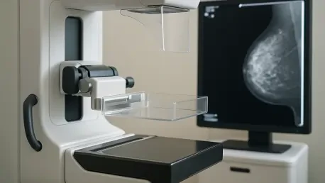
In the ever-evolving landscape of breast cancer screening, a transformative force is emerging through artificial intelligence (AI), poised to redefine how mammographic breast density is assessed. For decades, radiologists have relied on subjective visual evaluations to categorize breast density, a crucial indicator of breast cancer risk, using standardized systems like the American College of Radiology (ACR) Breast Imaging Reporting and Data System (BI-RADS). However, these human-led assessments often suffer from variability, leading to inconsistent risk stratification and screening recommendations that can impact patient outcomes. Could AI, with its data-driven precision, offer a long-sought solution to this persistent challenge? Imagine a technology that not only minimizes errors but also enhances the reliability of density classification, potentially reshaping the future of early detection. Recent studies are already unveiling promising results, as AI systems are tested to automate evaluations using full-field digital mammography (FFDM), hinting at a paradigm shift in clinical practice. The stakes couldn’t be higher—breast density directly influences mammographic sensitivity and cancer risk, often masking abnormalities in denser tissues and complicating diagnoses. This article delves deep into the heart of this innovation, exploring the potential for AI to overhaul breast density assessment by examining cutting-edge research and real-world implications. By shedding light on both the opportunities and obstacles, the discussion aims to uncover whether AI can truly elevate breast imaging to new heights of accuracy and personalization.
Why Breast Density Matters in Cancer Screening
Breast density, defined as the ratio of fibroglandular to fatty tissue in the breast, plays a pivotal role in breast cancer risk assessment and screening efficacy, with classifications ranging from A (least dense) to D (extremely dense) under the BI-RADS framework. Dense breasts, particularly those in categories C and D, are associated with an elevated risk of developing breast cancer, a connection well-documented in medical research. This heightened risk stems from the biological characteristics of dense tissue, which may foster an environment more conducive to tumor growth. Moreover, the presence of dense tissue complicates the interpretation of mammograms, as it can obscure potential abnormalities, leading to reduced diagnostic sensitivity. For women with dense breasts, the likelihood of missed diagnoses increases, posing a significant challenge to early detection efforts. Accurate classification of density is, therefore, not merely a technical exercise but a critical determinant of appropriate screening protocols and risk communication.
The prevalence of dense breasts adds another layer of urgency to the need for precise assessment, particularly among premenopausal women, where 24-35% are affected compared to 13-17% in postmenopausal groups. This demographic disparity underscores the importance of tailoring screening approaches based on density, often necessitating supplemental imaging like ultrasound or MRI for those at higher risk. Yet, the current reliance on visual evaluation by radiologists introduces inconsistencies that can undermine these efforts. Variability in assessments may result in some women receiving inadequate follow-up while others face unnecessary procedures, highlighting a gap in standardized care. AI emerges as a potential game-changer in this context, offering the promise of objective evaluations that could streamline decision-making and enhance patient outcomes by ensuring density is assessed with greater consistency and reliability.
Hurdles in Traditional Density Evaluation Methods
Traditional methods of assessing mammographic breast density hinge on radiologists’ visual interpretation of images, guided by the BI-RADS system, yet this approach is inherently subjective and prone to significant discrepancies. Even with standardized guidelines, two radiologists reviewing the same mammogram might assign different density categories, a phenomenon known as inter-observer variability. Such differences are not trivial; they can directly influence clinical decisions, potentially leading to misclassification of risk levels. For instance, a woman categorized as having less dense breasts by one reader might miss out on necessary additional screening if another reader deems the density higher. This lack of uniformity in assessment can erode trust in medical recommendations and create disparities in patient care.
Beyond discrepancies between different radiologists, intra-observer variability presents an additional challenge, where the same radiologist might classify a mammogram differently on separate occasions. Factors such as experience, fatigue, or subtle variations in image quality can contribute to these inconsistencies, further complicating the reliability of density evaluations. The implications extend to risk stratification and screening frequency, as inconsistent data can skew personalized care plans. In high-stakes environments like breast cancer screening, where early detection is paramount, these limitations underscore a critical need for a more dependable method. AI stands out as a promising avenue to address these issues, with the potential to automate assessments and provide a consistent benchmark that mitigates human error, paving the way for more uniform and effective screening protocols across diverse clinical settings.
AI’s Transformative Potential in Medical Imaging
Artificial intelligence, particularly through the application of deep learning algorithms, is carving out a transformative niche in medical imaging by offering unprecedented precision in analyzing complex data. These systems are meticulously trained on extensive datasets of annotated images to identify patterns and execute tasks such as detecting abnormalities or quantifying tissue characteristics with a level of accuracy that often rivals human expertise. In the realm of breast imaging, AI has already demonstrated substantial value in areas like lesion identification and optimizing clinical workflows. The ability to process vast amounts of imaging data rapidly positions AI as an indispensable asset in high-pressure environments, where balancing efficiency with diagnostic accuracy is a constant demand. This technological advancement hints at a future where routine tasks could be streamlined, allowing radiologists to focus on more intricate aspects of patient care.
When applied specifically to mammographic breast density assessment, AI brings a distinct advantage through its capacity for objectivity, sidestepping the biases and variability inherent in human evaluations. By employing sophisticated neural networks, these systems can isolate relevant breast tissue in mammograms, filtering out extraneous elements like pectoral muscle or imaging artifacts. The result is a precise calculation of fibroglandular versus fatty tissue proportions, which translates into both a categorical BI-RADS classification and a numeric density score. This dual functionality enhances the clinical relevance of AI outputs, providing radiologists with detailed insights that can refine risk assessments and inform tailored screening strategies. As AI continues to evolve, its integration into breast imaging holds the promise of shifting the field toward a more data-driven, personalized approach to cancer detection and prevention.
Evidence of AI’s Efficacy from Recent Studies
Groundbreaking research is shedding light on AI’s capability to revolutionize breast density assessment, with a notable study conducted at Kasr Alainy Hospital in Egypt offering compelling evidence of its potential. This retrospective analysis involved 592 female patients whose mammograms were evaluated using the Lunit INSIGHT FFDM software, alongside independent assessments by two radiologists with varying levels of experience. The findings revealed a remarkable alignment between AI and human judgment, with the system achieving an almost perfect agreement with one radiologist, reflected in a Cohen’s kappa value of 0.879 and a 92.4% agreement rate. Even with the second radiologist, where agreement was moderate at a kappa of 0.599 and a 73.6% rate, the results still underscored AI’s competitive performance. These outcomes suggest that AI could serve as a reliable tool for density classification, potentially surpassing the consistency of human evaluations in certain contexts.
A striking aspect of the study was the comparison between the radiologists themselves, which revealed only moderate agreement, with a kappa value of 0.552 and a 70.3% rate, particularly faltering in higher-density categories like C and D. This discrepancy highlights the entrenched issue of variability in visual assessments, a challenge that AI appears well-positioned to mitigate. Furthermore, the AI system provided numeric density scores that consistently correlated with BI-RADS categories, adding a quantitative layer to its assessments that could enhance clinical precision. Such capabilities indicate that AI might not only match human performance but also act as a stabilizing force in a field marked by inconsistency. The implications of these findings are profound, pointing toward a future where AI could standardize breast density readings, ensuring more uniform risk assessments and screening recommendations across diverse medical practices.
Clinical Benefits of Incorporating AI into Workflows
The integration of AI into mammographic workflows presents a transformative opportunity to elevate the standard of care in breast cancer screening by addressing longstanding issues of inconsistency in density assessment. By delivering objective evaluations, AI has the potential to significantly reduce variability among radiologists, ensuring that density classifications are uniform regardless of who interprets the mammogram. This consistency is crucial for effective risk communication, enabling healthcare providers to convey accurate information to patients about their breast cancer risk. For women identified with dense breasts, this could translate into more targeted screening plans, ensuring that those who require supplemental imaging, such as ultrasound or MRI, receive it promptly, while avoiding unnecessary procedures for others. The result is a more balanced approach to patient management that prioritizes both accuracy and efficiency.
In addition to improving individual care, AI’s automation capabilities offer substantial benefits in high-volume or resource-constrained clinical settings, where time and expertise are often stretched thin. By handling routine density assessments, AI can alleviate the workload on radiologists, allowing them to dedicate more attention to complex diagnostic challenges or direct patient interactions. This efficiency gain is particularly impactful in regions with limited access to specialized breast imaging professionals, where AI could bridge gaps in care delivery. Moreover, the standardization of density reporting through AI could enhance population-level data collection, providing richer datasets for research into breast cancer risk factors and screening outcomes. While hurdles such as implementation costs and training needs remain, the clinical advantages of AI in breast density assessment signal a promising step toward more precise, equitable, and effective screening practices that could ultimately save lives.
Pathways Forward for AI in Breast Imaging
While the initial research on AI’s role in breast density assessment paints an optimistic picture, significant work remains to ensure its widespread applicability and impact across diverse clinical landscapes. Single-center studies, though insightful, often lack the breadth to account for variations in patient demographics, imaging technologies, and healthcare practices globally. To address this, larger multicenter studies are essential to validate AI’s performance across different populations and equipment types. Such comprehensive research would help uncover any biases in algorithm training and ensure that AI tools deliver equitable benefits, regardless of geographic or socioeconomic context. Establishing this generalizability is a critical next step in building confidence in AI as a universal tool for breast density evaluation, capable of supporting radiologists in varied settings.
Another vital area for advancement lies in assessing AI’s influence on tangible clinical outcomes, beyond mere agreement rates with human assessments. Future studies should prioritize metrics like cancer detection rates, recall reductions, and overall improvements in patient prognosis to gauge the real-world value of AI-driven density assessments. Additionally, evaluating a range of AI platforms is necessary to determine whether performance differences arise from distinct algorithm designs or training datasets, which could inform best practices for clinical adoption. Practical considerations, including cost-effectiveness, radiologist training, and regulatory frameworks, must also be tackled to facilitate seamless integration into routine practice. By addressing these challenges, the path forward for AI in breast imaging can be paved with robust evidence and strategic planning, ensuring that its potential to transform mammographic breast density assessment is fully realized in the years ahead.


