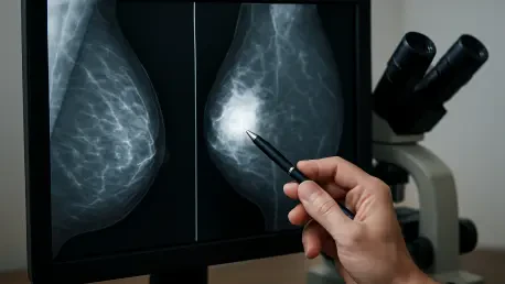

The ongoing quest for safer, more accurate breast cancer screening methods has led researchers to explore innovative alternatives to traditional mammography, which, despite its effectiveness, involves ionizing radiation. As the medical community increasingly seeks radiation-free options, the convergence of advanced imaging physics and sophisticated artificial intelligence is creating a new frontier in diagnostic capabilities. A significant breakthrough in this domain is now emerging, centering on the use of AI to automate and refine the complex processes behind novel imaging techniques. This development promises not only to enhance the precision of radiation-free scans but also to streamline clinical workflows, potentially making early and accurate detection more accessible. The integration of intelligent algorithms into technologies like acoustic computed tomography (CT) represents a pivotal step toward creating diagnostic tools that are both powerful and patient-friendly, signaling a potential paradigm shift in how breast health is monitored and managed.
The Intersection of AI and Advanced Imaging
A key development in this evolving landscape will be showcased at the prestigious SPIE Medical Imaging 2026 symposium, a major international event marking its 50th year in Vancouver, Canada. The conference, running from February 15–19, is a focal point for the latest advancements in medical imaging, with a strong emphasis on artificial intelligence and ultrasound technologies. At this event, Dr. Bilal Malik, Chief Science Officer for QT Imaging Holdings, Inc., is slated to deliver a featured oral presentation. His discussion will center on a significant innovation from the medical device company, which specializes in developing radiation-free solutions for breast health management. This presentation is anticipated to highlight the practical application of AI in enhancing the clinical utility of their cutting-edge imaging technology, offering a glimpse into the next generation of diagnostic tools that prioritize both patient safety and diagnostic accuracy by moving away from traditional radiation-based methods.
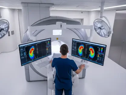
Dr. Malik’s presentation, titled “Automated breast boundary extraction from reflection data in breast acoustic CT,” will provide an in-depth look at a highly sophisticated, automated algorithm. This algorithm was meticulously developed by QT Imaging to precisely delineate the breast boundary in scans produced by its Breast Acoustic CT™ system. This technological leap is far more than a simple procedural improvement; it forms a critical piece of the company’s overarching strategy to fuse advanced, physics-based imaging with the power of modern artificial intelligence and machine learning (AI/ML). The goal of this synergy is to dramatically boost the clinical value and reliability of its non-invasive imaging technology. By automating what was once a complex and variable step, the algorithm establishes a consistent and accurate foundation upon which all subsequent image processing and analysis can be built, ensuring higher quality results and paving the way for more complex AI-driven applications in the near future.
Foundational Algorithms for Future Clinical Tools
The immediate impact of this automated boundary detection algorithm is profound, as it serves as the foundational element for the entire imaging pipeline. Its precise function is essential for significantly improving the accuracy of image reconstructions, which is the process of creating a detailed picture from the raw scan data. Furthermore, the algorithm is instrumental in determining the correct gain factor for reflection imaging, a key parameter that affects image brightness and contrast. It also enables more effective denoising, filtering out irrelevant signals to produce a clearer and more interpretable image. Crucially, this automated boundary is a prerequisite for the accurate calculation of breast density, a vital biomarker for assessing breast cancer risk. Dr. Malik has emphasized that the multi-step algorithm is not only fast and efficient but has also been rigorously validated on an extensive dataset and is already fully integrated into the company’s production software, underscoring its readiness for widespread clinical use.
This validated algorithm represented more than just an enhancement to an existing system; it was a cornerstone upon which future AI and machine learning initiatives could be built. The successful automation of boundary detection provided the clean, reliable data necessary for developing more advanced diagnostic aids. These future tools were aimed at tackling complex challenges such as automated lesion segmentation, where the AI would be trained to identify and outline suspicious areas on its own. The ultimate vision was to create comprehensive clinical decision support systems. Such systems would not only assist radiologists by improving lesion characterization but also streamline entire clinical workflows, reducing the time from scan to diagnosis. By laying this fundamental groundwork, the company had positioned itself to pioneer new applications of AI that could potentially lead to earlier cancer detection and more personalized patient care pathways.