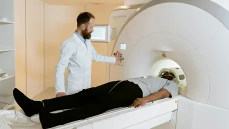

A diagnosis of a high-grade glioma, one of the most aggressive forms of brain cancer, plunges patients and their families into a world of clinical uncertainty where effective treatment strategies are desperately needed. The standard approach to understanding these tumors involves an invasive surgical biopsy, a procedure that carries significant risks and often provides an incomplete picture due to the complex, heterogeneous nature of the cancer. However, recent advancements at the intersection of artificial intelligence and medical imaging are beginning to challenge this paradigm, offering a glimpse into a future where a simple MRI scan could non-invasively unlock a tumor’s biological secrets and guide physicians toward the most effective personalized therapies, fundamentally changing the landscape of neuro-oncology. This technological leap promises not only to enhance patient safety by avoiding surgery but also to provide a far more comprehensive assessment of the tumor, paving the way for a new era of precision medicine in the fight against this devastating disease.
A New Alliance AI Radiomics and MRI
Decoding Tumors with Digital Fingerprints
The foundation of this innovative approach rests within the field of radiomics, a sophisticated discipline that transforms conventional medical images into a vast repository of high-dimensional, mineable data. For this research, scientists leveraged contrast-enhanced Magnetic Resonance Imaging (MRI), a standard clinical tool renowned for its ability to visualize the intricate and varied structures within gliomas. Using advanced data-characterization algorithms, a multitude of quantitative features were extracted from these scans, encompassing subtle variations in texture, complex shape descriptors, and nuanced intensity distributions. These radiomic features, which are entirely imperceptible to the human eye, function as a unique digital fingerprint for the tumor. This process effectively converts a visual diagnostic image into a rich, quantitative dataset that captures the underlying biological environment of the cancer, including critical information about its cellularity, vascularity, and areas of necrosis, all without a single incision.
This immense and intricate dataset of digital fingerprints was then fed into a highly sophisticated machine learning model. The researchers specifically selected a gradient boosting algorithm, a powerful and robust technique celebrated for its exceptional predictive accuracy when handling complex, high-dimensional information. The algorithm was meticulously trained to perform a task far beyond human capability: to identify and learn the intricate patterns and hidden correlations between the thousands of radiomic features and the ground-truth biological data. In essence, the artificial intelligence was taught to “read” the subtle language of the MRI scans and translate the tumor’s digital signature into a clear, actionable biological insight. This computational process represents a paradigm shift, moving from subjective visual interpretation to objective, data-driven analysis that can decode the molecular makeup of a tumor directly from standard clinical imaging, opening up unprecedented diagnostic possibilities.
The EGFR Biomarker a Key to Unlocking Treatment
The AI model was specifically engineered to predict the expression level of a critical molecule: the Epidermal Growth Factor Receptor (EGFR). Within the context of high-grade gliomas, EGFR is not merely a passive cellular component; it is a primary driver of malignancy. Elevated levels of EGFR expression are strongly correlated with increased tumor aggressiveness, rapid proliferation, and consequently, a poorer prognosis for the patient. Its pivotal role in tumor progression has made it a vital diagnostic biomarker and a prime target for a new generation of precision therapies, including highly specific antibody-drug conjugates designed to seek out and destroy cancer cells overexpressing this receptor. Therefore, the ability to accurately and reliably determine a tumor’s EGFR status is paramount. It serves as a crucial fork in the road for clinical decision-making, dictating whether a patient is a suitable candidate for these advanced, targeted treatments or if an alternative therapeutic strategy should be pursued.
This AI-driven approach provides a powerful solution to one of the most significant hurdles in glioma treatment. The current gold standard for assessing EGFR status is the surgical biopsy, an invasive procedure that is not without risk of complications and patient discomfort. More critically, a biopsy only samples a tiny fraction of the tumor, a major limitation given that gliomas are notoriously heterogeneous. This means different regions of the same tumor can exhibit vastly different molecular characteristics, and a single sample may fail to capture the complete biological profile, leading to a misinformed treatment plan. The radiomics model circumvents this problem entirely by analyzing the entire tumor volume captured in the MRI scan. This non-invasive, holistic assessment provides a more comprehensive and representative picture of the tumor’s EGFR expression, empowering clinicians to make more informed, personalized treatment decisions and significantly improving the odds of matching the right patient to the right therapy.
Proving the Concept and Paving the Future
Validating the Model with Advanced Organoids
A critical step in establishing the credibility and potential of this predictive model was its rigorous validation process. To achieve this, the researchers utilized high-grade glioma organoid models, which represent a significant leap forward in preclinical research. These are not simple, two-dimensional cell cultures; they are sophisticated, three-dimensional structures meticulously grown in a laboratory environment to mirror the complex architecture, cellular diversity, and biological behavior of actual human tumors. By testing and confirming the AI model’s predictive accuracy on these scientifically robust and ethically viable platforms, the study ensured its findings were grounded in a system that closely approximates real-world clinical conditions. This meticulous preclinical validation is an indispensable milestone, providing the strong evidence needed to build confidence in the model’s reliability and pave the way for its eventual translation into a tool used for human patients.
The use of organoids was essential for demonstrating that the model’s predictions were not just statistical correlations but were rooted in genuine biological phenomena. Unlike simpler experimental setups, these advanced models successfully replicate key aspects of tumor biology, including cell-to-cell interactions and the development of a complex microenvironment. Validating the radiomics-based predictions against the known EGFR status of these organoids confirmed that the digital fingerprints extracted from MRI scans truly reflect the underlying molecular reality of the tumor. This successful demonstration in a highly relevant preclinical setting is a crucial proof of concept, showing that the AI can indeed bridge the gap between medical imaging and molecular diagnostics. It strengthens the case for advancing this technology into larger-scale human clinical trials, moving it one step closer to becoming a transformative tool in the clinical management of glioma.
A New Standard in Patient Care
The successful development and validation of this radiomics-based model had profound implications for the future management of glioma. Its demonstrated ability to non-invasively predict a key biomarker offered a safer, more comfortable, and potentially more accurate alternative to the inherent risks and limitations of surgical biopsies. By furnishing clinicians with a detailed assessment of a tumor’s biology before treatment even commenced, the model empowered them to make more informed and personalized decisions. This capability perfectly aligned with the central tenets of precision medicine: moving away from a one-size-fits-all approach and instead tailoring therapeutic protocols to the unique molecular characteristics of an individual patient’s cancer. Such targeted strategies were expected to significantly enhance therapeutic efficacy while simultaneously minimizing the adverse side effects often associated with treatments that were not well-matched to the tumor’s specific biology.
Furthermore, this pioneering research contributed significantly to the rapidly advancing field of artificial intelligence in healthcare. It provided a compelling and concrete example of how machine learning could transform standard clinical data, such as an MRI scan, into a powerful tool for predictive diagnostics. As this technology continued to mature, it promised to streamline clinical workflows, reduce diagnostic uncertainty, and ultimately set a new benchmark for the standard of patient care. The work by Tan et al. was not seen as an endpoint but as a significant milestone on a longer journey toward more effective cancer treatment. The path forward involved refining the model and validating its performance in larger, multi-center human clinical trials, with the ultimate goal of integrating these advanced computational tools into everyday oncology. This study illuminated a promising new frontier where the synergy of advanced imaging and AI brought the vision of a more precise, patient-centered approach to reality.