A groundbreaking advance in medical imaging promises to redefine early disease detection, drawing significant attention to the work conducted at UC Davis Health. With the deployment of Explorer, a state-of-the-art total body scanner, researchers have targeted the permeability of the blood-brain barrier (BBB) using positron emission tomography (PET). This revolutionary application could potentially lead to unprecedented improvements in diagnosing neurological conditions early in their progression. Key contributors include postdoctoral fellow Kevin Chung and radiology experts such as Guobao Wang, Simon Cherry, and Ramsey Badawi. These figures are championing efforts to bridge the gap between complex scientific inquiry and tangible healthcare outcomes, setting a new benchmark in the assessment and understanding of systemic health conditions that affect the brain.
Innovations in Blood-Brain Barrier Analysis
The recent study conducted at UC Davis Health unlocks new insights into the blood-brain barrier (BBB)’s permeability, an essential function safeguarding the brain from harmful entities, while highlighting its role in the early detection of brain diseases. Traditional methodologies, like magnetic resonance imaging (MRI), generally focus on detecting permeability changes only in advanced disease stages, often missing initial alterations. The introduction of positron emission tomography (PET) into this area of research marks a pivotal shift, as it offers an innovative solution to identifying earlier signs of BBB dysfunction. PET imaging’s ability to non-invasively evaluate changes that precede overt symptoms offers collaborating scientists an invaluable tool to enhance their understanding of BBB dynamics. By overcoming prior limitations in identifying early indicators, PET imaging paves the way for preventive strategies, promising to tackle neurological conditions before they advance significantly.
Moreover, understanding the permeability of the BBB extends beyond just early disease detection. It opens doors to explore how various systemic conditions, such as cancer or neurodegenerative diseases, impact brain health. This pioneering step pushes the envelope by not only analyzing the BBB’s protective role but also observing its variations due to several external factors and systemic disorders. The capacity of PET imaging to monitor such detailed changes underscores the potential for new therapeutic targets and intervention points. The shift from merely observing symptoms to understanding underlying causes aligns with an overarching aim in medical research: improving patient outcomes through timely, accurate diagnoses that inform effective treatment regimes. As research progresses, this innovative approach stands to contribute significantly to broadening the scope and depth of knowledge concerning brain barriers and their pertinent implications.
Explorer’s Enhanced Imaging Capabilities
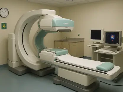
The Explorer total body scanner, a brainchild of experts Simon Cherry and Ramsey Badawi, developed with support from the National Institutes of Health, heralds a new era of PET imaging by introducing unparalleled capabilities. Unlike conventional imaging devices, Explorer offers a remarkable advancement in temporal resolution, allowing researchers to observe rapid biological transformations almost in real time. With the capability to produce imaging frames that are as quick as one second, scientists can capture subtle physiological changes, such as those occurring in vital transporters across the BBB that other imaging technologies often miss. This swift data acquisition provides a significant edge in biomedical research, facilitating earlier and more precise detections of potential alterations in brain barrier mechanisms.
Integral to the high efficacy of Explorer is the sophisticated mathematical modeling that supplements its imaging precision. This advanced modeling accurately interprets the vast volumes of data generated, unveiling even the faintest signals of BBB transformation. Through this combination of cutting-edge imaging and analytics, Explorer can detect nuanced variations that could signal the onset of neurologically compromising conditions. These tools offer researchers a detailed panorama of BBB health that surpasses traditional methodologies, equipping them with the insights needed to proactively address emerging medical challenges. Explorer represents a groundbreaking confluence of technological innovation and scientific inquiry, augmenting the ability to detect and diagnose brain-related disorders with unprecedented accuracy and foresight.
Extensive Versatility of Radiotracers
One notable outcome of the UC Davis study is the demonstration of PET imaging’s extensive versatility through the employment of over a thousand existing radiotracers. This remarkable adaptability not only extends the capabilities of such imaging but also broadens its applicability across diverse medical contexts. Each radiotracer presents unique interaction properties with the body’s biological processes, allowing researchers to target specific pathways and assess BBB permeability under various conditions. This comprehensive approach deepens the understanding of how normal aging, environmental factors, or specific health conditions influence the brain’s defenses. By using this vast array of radiotracers, the researchers can customize imaging procedures to answer specific clinical and research questions, thus pushing the boundaries of current diagnostic practices to new heights.
Additionally, PET imaging’s ability to adapt to changes in BBB permeability expands its utility in monitoring responses to external stressors and diseases, leading to improved diagnostic precision. The flexible nature of this imaging modality enables researchers to tailor it according to specific study requirements or medical inquiries, whether tracking liver inflammation’s impact on the BBB or mapping aging’s effects. This dynamic capability further solidifies PET imaging’s role as a cornerstone in the landscape of modern medical diagnostics. As researchers continue to explore this adaptability, the potential for PET to transform conventional diagnostic and therapeutic approaches amplifies, providing hope for effective management and intervention strategies in preventing and treating various neurological conditions.
Collaboration and Support Framework
The success of this innovative research at UC Davis Health is attributed to a robust collaboration involving an interdisciplinary team of researchers and aligning with substantial institutional support. Prominent contributors such as Yasser Abdelhafez, Benjamin Spencer, and several others brought their expertise to the forefront, facilitating significant strides in imaging technologies. This collective effort underscores the critical role of teamwork in advancing scientific endeavors, ensuring a broad spectrum of insights and meticulous attention to each aspect of study design and execution. Their combined expertise not only enhanced the research’s quality but also ensured that all findings were meticulously validated, providing a strong foundation for future studies in medical imaging.
Institutional backing from esteemed bodies, including the National Institute of Biomedical Imaging and Bioengineering (NIBIB), National Cancer Institute (NCI), and National Institute of Diabetes and Digestive and Kidney Diseases (NIDDK), further bolstered this project’s impact and reach. Their support signifies a broader acknowledgment of the potential benefits this breakthrough offers, not just within the field of diagnostic imaging but across a myriad of systemic health concerns. Such investments highlight a commitment to pioneering research, fostering innovation that promises significant advancements in predictive medicine. This collaborative milieu serves as a testament to what can be achieved when academia, industry, and healthcare institutions unite towards a common goal: the elevation of medical practice standards and the betterment of patient outcomes worldwide.
Towards Early Diagnosis and Intervention
A recent study at UC Davis Health presents new insights into the permeability of the blood-brain barrier (BBB), crucial for protecting the brain from harmful substances, and emphasizes its role in the early detection of brain diseases. Typical methods such as MRI often focus on identifying permeability changes in advanced stages, overlooking initial variations. However, the integration of positron emission tomography (PET) signifies a significant change, providing a novel way to spot earlier signs of BBB dysfunction. PET imaging can non-invasively assess changes before obvious symptoms appear, offering researchers an invaluable tool to deepen their understanding of BBB dynamics. This method helps overcome previous limitations in detecting early signs, paving the way for preventive strategies against advancing neurological disorders. Understanding BBB permeability also reveals how systemic conditions like cancer or neurodegenerative diseases affect brain health. PET imaging’s capability to monitor these changes underscores its potential for uncovering new therapeutic targets, aiding in timely, accurate diagnoses, and improving patient outcomes as research progresses.


