Imagine a scenario in a bustling hospital where a child struggles to breathe, and doctors are racing against time to uncover the cause—a tiny piece of food, invisible on a chest scan, lodged deep in the airway, posing a silent but deadly threat that could lead to severe complications. This is the daunting reality of radiolucent foreign body aspiration (FBA), a condition where small objects evade detection due to their transparency on imaging, often leading to delayed diagnoses and severe issues like infections or blockages. For years, even the most skilled radiologists have grappled with alarmingly low detection rates, highlighting a critical gap in medical diagnostics. Now, a revolutionary breakthrough offers a beacon of hope. Researchers from the University of Southampton, working alongside clinical partners in Wuhan, China, have unveiled an AI-powered tool that dramatically outperforms human experts in spotting these hidden dangers. Published in npj Digital Medicine, this innovation marks a turning point, promising to reshape how respiratory challenges are addressed in clinical settings.
The Challenge of Hidden Threats in Chest Scans
Addressing the Invisible Danger
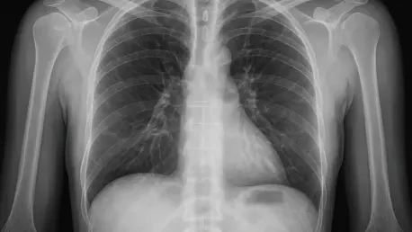
Detecting small foreign objects in the airways remains one of the most perplexing challenges in medical imaging, as these items often blend seamlessly into surrounding tissues on chest CT scans. Known as radiolucent foreign body aspiration, this condition frequently involves organic materials like food particles that do not appear distinctly on standard imaging, making them nearly impossible to identify without invasive procedures. The consequences of missing such objects can be dire, ranging from persistent respiratory distress to life-threatening airway obstructions or recurrent infections. Even seasoned radiologists struggle with this task due to the subtle nature of the anomalies, often resulting in delayed interventions. The urgency to improve detection methods is driven by the need to protect vulnerable patients, particularly children, who are at higher risk of such incidents. This diagnostic blind spot has long underscored the limitations of human perception in complex imaging scenarios, setting the stage for technological intervention.
Beyond the immediate health risks, the broader impact of undetected foreign bodies extends to long-term complications that burden both patients and healthcare systems. Chronic lung damage, repeated hospital visits, and the emotional toll on families are just some of the ripple effects when diagnoses are missed. Traditional imaging techniques, while valuable for many conditions, fall short in sensitivity for these specific cases, often prioritizing the avoidance of false positives over catching every potential threat. This cautious approach, though well-intentioned, leaves many cases undiagnosed until symptoms worsen significantly. The frustration among clinicians is palpable, as they lack the tools to confidently identify these hidden dangers without resorting to more invasive methods like bronchoscopy, which carries its own risks and costs. Addressing this invisible danger requires a paradigm shift, one that leverages cutting-edge solutions to bridge the gap between what the human eye can see and what remains hidden in the intricate landscape of the human airway.
Limitations of Traditional Methods
Conventional diagnostic approaches to chest imaging, while precise in minimizing false positives, suffer from a critical flaw: their low sensitivity in detecting radiolucent objects. Radiologists, relying on standard CT scans, achieve a detection rate of only 36% in confirmed cases of foreign body aspiration, a statistic that reveals the profound challenge of identifying anomalies that do not contrast sharply with surrounding tissues. This gap often means that patients endure prolonged uncertainty, with symptoms misattributed to other conditions until more invasive tests are conducted. The stakes are high, as delayed diagnosis can escalate minor issues into emergencies requiring urgent intervention. The inherent limitations of human visual analysis in such nuanced scenarios highlight why traditional methods, despite their strengths in other areas, are insufficient for this specific medical hurdle, pushing the field toward innovative alternatives.
Moreover, the reliance on human expertise alone places immense pressure on radiologists, who must navigate the fine line between caution and accuracy under time constraints. The risk of overlooking a foreign body is compounded by the variability in experience levels among clinicians, as well as the sheer volume of scans they must review daily. Even with advanced training, the human eye struggles to discern the faint textural differences that might indicate a hidden object, often leading to missed opportunities for early treatment. This systemic challenge is not just a technical issue but a call to action for tools that can enhance diagnostic precision without overwhelming medical professionals. The persistent shortcomings of current methods have fueled a growing consensus in the medical community that supplementary technologies are essential to address this diagnostic blind spot, paving the way for solutions that can redefine the standard of care in respiratory medicine.
AI as a Diagnostic Game-Changer
Unveiling Superior Detection Capabilities
In a remarkable stride forward, an AI-powered diagnostic tool developed by researchers has demonstrated an extraordinary ability to detect radiolucent foreign bodies in chest CT scans, achieving a sensitivity rate of 71% compared to a mere 36% for experienced radiologists. This significant disparity underscores the technology’s potential to transform how hidden threats are identified in clinical settings. Evaluated against a set of 70 scans, including confirmed cases validated through bronchoscopy, the AI not only excelled in spotting elusive objects but also maintained a balanced diagnostic performance with a precision of 77% and an F1 score of 74%, far outpacing human scores. This capability to flag potential issues with greater accuracy offers a lifeline in situations where time is critical, reducing the likelihood of missed diagnoses. The implications of such performance are profound, signaling a shift toward more reliable detection methods in respiratory care.
Equally striking is the AI’s capacity to handle the subtleties of chest imaging that often confound human observers, providing a level of consistency that is difficult to achieve manually. By focusing on minute textural and morphological differences, the system identifies anomalies that blend into the complex anatomy of the airways, a task that challenges even the most trained professionals. This superior detection rate does not just represent a numerical advantage but translates into real-world benefits, such as earlier interventions that can prevent complications like airway blockages or chronic infections. While the tool does produce some false positives, its overall balance ensures that clinicians are not inundated with unnecessary alerts, maintaining a practical approach to diagnostics. This advancement stands as a testament to how technology can address longstanding gaps in medical imaging, offering a new benchmark for accuracy in identifying life-threatening conditions.
Technical Innovation Behind the Tool
At the heart of this groundbreaking AI tool lies a sophisticated deep learning model that integrates an airway segmentation framework known as MedpSeg with a convolutional neural network (CNN), enabling unparalleled precision in chest CT analysis. Trained on a comprehensive dataset of over 400 scans, the system has learned to map the intricate bronchial architecture and pinpoint suspicious regions that might harbor foreign bodies. This technical prowess allows it to detect subtle patterns and anomalies that are imperceptible to the human eye, such as faint density shifts or irregular textures within the airways. The meticulous design of the model ensures that it not only identifies potential threats but also contextualizes them within the anatomical structure, providing clinicians with actionable insights. Such innovation represents a significant leap in diagnostic technology, pushing the boundaries of what is possible in medical imaging through computational intelligence.
The development process of this AI system also reflects a commitment to robustness, as it was rigorously tested to ensure reliability across varied imaging conditions and patient profiles. By leveraging neural networks, the tool continuously refines its understanding of complex scan data, adapting to the nuances of individual cases with a level of detail that manual analysis cannot match. This adaptability is key to its success, allowing the system to maintain high performance even when faced with challenging or atypical scans. Furthermore, the integration of airway segmentation ensures that the AI focuses on the most relevant areas, minimizing irrelevant noise and enhancing diagnostic clarity. This technical foundation not only explains the tool’s superior detection capabilities but also highlights the potential for similar AI-driven solutions to tackle other intricate diagnostic challenges in medicine, setting a precedent for future innovations in healthcare technology.
A Collaborative Future for Medicine
AI as a Partner, Not a Replacement
Far from aiming to supplant radiologists, this AI diagnostic tool is envisioned as a vital “second observer,” designed to augment human expertise in the complex landscape of medical imaging. By acting as a supportive partner, the system highlights subtle anomalies that might escape even the most diligent professionals, thereby reducing diagnostic uncertainty in challenging cases. This collaborative approach aligns with the concept of augmented intelligence, where technology enhances rather than competes with clinical judgment. Lead researcher Dr. Yihua Wang has emphasized that the goal is to empower radiologists with a reliable ally, ensuring that critical findings are not overlooked amid high workloads or intricate scans. Such synergy between human skill and computational precision holds the promise of elevating the standard of care, particularly in specialized fields like respiratory diagnostics where accuracy is paramount.
This partnership model also addresses concerns about the dehumanization of medicine by reinforcing the irreplaceable value of clinical experience and empathy. The AI tool serves as a complementary resource, providing radiologists with additional confidence in their assessments while allowing them to focus on patient interaction and holistic care. Its role is to handle the heavy lifting of data analysis, sifting through countless scan details to flag potential issues for further review, thus alleviating some of the cognitive burden on medical staff. This balance ensures that technology remains a servant to human expertise, not a substitute, fostering trust among healthcare providers who may be wary of over-reliance on automated systems. By integrating seamlessly into existing workflows, the AI paves the way for a future where diagnostics are both more precise and more human-centered, blending the best of both worlds to improve patient outcomes.
Streamlining Clinical Workflows
One of the most promising aspects of this AI tool is its potential to streamline clinical workflows, enabling faster and more accurate treatment decisions in high-stakes environments. By rapidly identifying potential foreign bodies in chest scans, the system cuts down the time spent on diagnostic uncertainty, allowing clinicians to prioritize urgent cases and initiate interventions sooner. This efficiency is particularly crucial in emergency settings, where every minute can make a difference in preventing severe complications like airway obstructions or infections. The tool’s ability to deliver consistent results also reduces the need for repeated scans or invasive follow-ups, saving valuable resources and minimizing patient discomfort. Such improvements in workflow not only enhance the quality of care but also alleviate pressure on overburdened healthcare systems facing growing demands.
Additionally, the integration of this AI into routine practice could reshape how radiologists allocate their focus, freeing them to tackle more complex interpretive tasks while the system handles initial screenings. This redistribution of effort ensures that human expertise is applied where it is most needed, such as in confirming AI findings or addressing ambiguous cases that require nuanced judgment. The ripple effect of streamlined processes extends beyond individual patient encounters, potentially reducing wait times and improving overall hospital efficiency. As healthcare facilities grapple with increasing caseloads, tools like this offer a scalable solution to enhance throughput without compromising accuracy. The vision of a more agile diagnostic pipeline, supported by technology, underscores the transformative impact this innovation could have on the day-to-day operations of medical institutions, ultimately benefiting both providers and patients alike.
Looking Ahead to Broader Impact
Plans for Validation and Equity
As this AI diagnostic tool gains traction, the research team is keenly focused on expanding its validation through larger, multicenter trials to ensure its reliability across diverse clinical settings. These studies aim to refine the model’s accuracy by exposing it to a wider range of patient demographics and imaging conditions, addressing any potential gaps in performance. A critical aspect of this phase is tackling algorithmic biases that could skew results in specific populations, ensuring that the technology delivers equitable outcomes regardless of age, ethnicity, or geographic location. This commitment to fairness reflects a broader understanding in the medical community that diagnostic tools must be inclusive to be truly effective. By prioritizing rigorous testing, the researchers are laying the groundwork for a solution that can be trusted by clinicians worldwide, strengthening its case for eventual regulatory approval and clinical integration.
Equally important is the emphasis on collaboration during this validation process, as partnerships with international medical institutions provide access to varied datasets and expertise. These alliances help in fine-tuning the AI to account for anatomical variations and cultural differences in healthcare practices, which might influence diagnostic needs. The goal is to create a tool that is not only precise but also adaptable, capable of serving both high-resource hospitals and under-equipped facilities with equal efficacy. This focus on equity extends to addressing ethical concerns, such as ensuring patient data privacy during testing and deployment. By taking these steps, the team aims to build confidence among stakeholders that the technology is both scientifically sound and socially responsible, paving the way for its acceptance as a standard diagnostic aid in respiratory medicine across the globe.
Potential for Widespread Adoption
The vision of integrating this AI tool into routine clinical practice is gaining momentum, fueled by the promise of revolutionizing respiratory diagnostics on a global scale. Supported by international collaboration and funding from entities like the UK Medical Research Council, the project exemplifies how collective efforts can drive innovation from research labs to hospital rooms. Widespread adoption could mean a seismic shift in how foreign body aspiration cases are managed, with earlier detections leading to fewer complications and improved survival rates. The scalability of the technology also makes it a viable option for diverse healthcare environments, from urban medical centers to rural clinics, potentially bridging disparities in access to advanced diagnostics. This prospect of universal reach amplifies the tool’s impact, positioning it as a cornerstone of modern medical practice.
Looking back, the journey of this AI system reflects a relentless pursuit of precision, as researchers tirelessly refined its capabilities to outshine traditional methods in past evaluations. The success in controlled studies, where it consistently surpassed human detection rates, cemented its potential as a transformative force. Beyond individual patient care, the adoption of such tools in the past hinted at a future where healthcare systems could operate with greater efficiency, reducing costs associated with delayed treatments and extended hospital stays. Reflecting on those early achievements, it became clear that the path forward involves not just technological refinement but also strategic planning to ensure seamless integration into existing infrastructures. The next steps should focus on training programs for clinicians, robust support systems for implementation, and continuous updates to the AI model to keep pace with evolving medical challenges, ensuring that past successes translate into lasting improvements for patient care worldwide.
